 Movie
Movie Controller


+ Open data

- Basic information
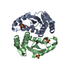
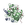
| Entry | Database: PDB / ID: 1j6w | ||||||
|---|---|---|---|---|---|---|---|
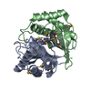
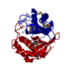
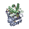
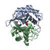
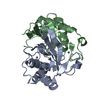
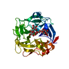
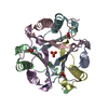
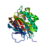
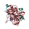
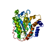
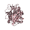
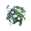
| Title | CRYSTAL STRUCTURE OF HAEMOPHILUS INFLUENZAE LUXS | ||||||
 Components Components | AUTOINDUCER-2 PRODUCTION PROTEIN LUXS | ||||||
 Keywords Keywords | SIGNALING PROTEIN / alpha-beta fold | ||||||
| Function / homology |  Function and homology information Function and homology informationS-ribosylhomocysteine lyase / S-ribosylhomocysteine lyase activity / : / quorum sensing / iron ion binding / cytosol Similarity search - Function | ||||||
| Biological species |  Haemophilus influenzae (bacteria) Haemophilus influenzae (bacteria) | ||||||
| Method |  X-RAY DIFFRACTION / SYNCHROTRON / MAD / Resolution: 2.1 Å X-RAY DIFFRACTION / SYNCHROTRON / MAD / Resolution: 2.1 Å | ||||||
 Authors Authors | Lewis, H.A. / Furlong, E.B. / Bergseid, M.G. / Sanderson, W.E. / Buchanan, S.G. | ||||||
 Citation Citation | Journal: Structure / Year: 2001 Title: A structural genomics approach to the study of quorum sensing: crystal structures of three LuxS orthologs. Authors: Lewis, H.A. / Furlong, E.B. / Laubert, B. / Eroshkina, G.A. / Batiyenko, Y. / Adams, J.M. / Bergseid, M.G. / Marsh, C.D. / Peat, T.S. / Sanderson, W.E. / Sauder, J.M. / Buchanan, S.G. #1: Journal: Proteins / Year: 2005Title: Structural analysis of a set of proteins resulting from a bacterial genomics project Authors: Badger, J. / Sauder, J.M. / Adams, J.M. / Antonysamy, S. / Bain, K. / Bergseid, M.G. / Buchanan, S.G. / Buchanan, M.D. / Batiyenko, Y. / Christopher, J.A. / Emtage, S. / Eroshkina, A. / ...Authors: Badger, J. / Sauder, J.M. / Adams, J.M. / Antonysamy, S. / Bain, K. / Bergseid, M.G. / Buchanan, S.G. / Buchanan, M.D. / Batiyenko, Y. / Christopher, J.A. / Emtage, S. / Eroshkina, A. / Feil, I. / Furlong, E.B. / Gajiwala, K.S. / Gao, X. / He, D. / Hendle, J. / Huber, A. / Hoda, K. / Kearins, P. / Kissinger, C. / Laubert, B. / Lewis, H.A. / Lin, J. / Loomis, K. / Lorimer, D. / Louie, G. / Maletic, M. / Marsh, C.D. / Miller, I. / Molinari, J. / Muller-Dieckmann, H.J. / Newman, J.M. / Noland, B.W. / Pagarigan, B. / Park, F. / Peat, T.S. / Post, K.W. / Radojicic, S. / Ramos, A. / Romero, R. / Rutter, M.E. / Sanderson, W.E. / Schwinn, K.D. / Tresser, J. / Winhoven, J. / Wright, T.A. / Wu, L. / Xu, J. / Harris, T.J. | ||||||
| History |
|
- Structure visualization
| Structure viewer | Molecule: MolmilJmol/JSmol |
|---|
- Downloads & links
-Download
| PDBx/mmCIF format | 1j6w.cif.gz | 77 KB | Display | PDBx/mmCIF format |
|---|---|---|---|---|
| PDB format | pdb1j6w.ent.gz | 58.3 KB | Display | PDB format |
| PDBx/mmJSON format | 1j6w.json.gz | Tree view | PDBx/mmJSON format | |
| Others |  Other downloads Other downloads |
-Validation report
| Arichive directory | https://data.pdbj.org/pub/pdb/validation_reports/j6/1j6wftp://data.pdbj.org/pub/pdb/validation_reports/j6/1j6w | HTTPS FTP |
|---|
-Related structure data
-Links
 PDBj
PDBj

- Assembly
| Deposited unit | 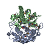
| ||||||||||
|---|---|---|---|---|---|---|---|---|---|---|---|
| 1 |
| ||||||||||
| Unit cell |
|
-Components
| #1: Protein | Mass: 19849.512 Da / Num. of mol.: 2 Source method: isolated from a genetically manipulated source Source: (gene. exp.) Haemophilus influenzae (bacteria) / Production host: #2: Chemical |   Mass: 65.409 Da / Num. of mol.: 2 / Source method: obtained synthetically / Formula: Zn Mass: 65.409 Da / Num. of mol.: 2 / Source method: obtained synthetically / Formula: Zn#3: Chemical |   Type: L-peptide linking / Mass: 149.211 Da / Num. of mol.: 2 / Source method: obtained synthetically / Formula: C5H11NO2S Type: L-peptide linking / Mass: 149.211 Da / Num. of mol.: 2 / Source method: obtained synthetically / Formula: C5H11NO2S#4: Water | ChemComp-HOH / |  Mass: 18.015 Da / Num. of mol.: 57 / Source method: isolated from a natural source / Formula: H2O Mass: 18.015 Da / Num. of mol.: 57 / Source method: isolated from a natural source / Formula: H2OHas protein modification | Y | |
|---|
-Experimental details
-Experiment
| Experiment | Method: X-RAY DIFFRACTION / Number of used crystals: 1 |
|---|
- Sample preparation
| Crystal | Density Matthews: 3.14 Å3/Da / Density % sol: 60.5 % | |||||||||||||||||||||||||
|---|---|---|---|---|---|---|---|---|---|---|---|---|---|---|---|---|---|---|---|---|---|---|---|---|---|---|
| Crystal grow | Temperature: 285 K / Method: vapor diffusion, hanging drop / pH: 6.25 Details: PEG MME 5000, MES, BME, NaCl, pH 6.25, VAPOR DIFFUSION, HANGING DROP at 285K, temperature 285.0K | |||||||||||||||||||||||||
| Crystal grow | *PLUS Temperature: 12 ℃ | |||||||||||||||||||||||||
| Components of the solutions | *PLUS
|
-Data collection
| Diffraction | Mean temperature: 100 K | |||||||||
|---|---|---|---|---|---|---|---|---|---|---|
| Diffraction source | Source: SYNCHROTRON / Site: APS  / Beamline: 32-ID / Wavelength: 0.9795, 0.9641 / Beamline: 32-ID / Wavelength: 0.9795, 0.9641 | |||||||||
| Detector | Type: MARRESEARCH / Detector: CCD / Date: Sep 5, 2000 | |||||||||
| Radiation | Monochromator: Graphite / Protocol: MAD / Monochromatic (M) / Laue (L): M / Scattering type: x-ray | |||||||||
| Radiation wavelength |
| |||||||||
| Reflection | Resolution: 2.1→32 Å / Num. all: 50926 / Num. obs: 50722 / % possible obs: 99.6 % / Observed criterion σ(F): 0 / Observed criterion σ(I): 0 / Redundancy: 7.2 % / Biso Wilson estimate: 25.9 Å2 / Rmerge(I) obs: 0.064 / Net I/σ(I): 24.6 | |||||||||
| Reflection shell | Resolution: 2.1→2.18 Å / Redundancy: 3.7 % / Rmerge(I) obs: 0.094 / % possible all: 100 | |||||||||
| Reflection shell | *PLUS % possible obs: 98.6 % / Rmerge(I) obs: 0.184 |
- Processing
| Software |
| |||||||||||||||||||||||||
|---|---|---|---|---|---|---|---|---|---|---|---|---|---|---|---|---|---|---|---|---|---|---|---|---|---|---|
| Refinement | Method to determine structure: MAD / Resolution: 2.1→30 Å / σ(F): 0 / σ(I): 0 / Stereochemistry target values: Engh & Huber
| |||||||||||||||||||||||||
| Refinement step | Cycle: LAST / Resolution: 2.1→30 Å
| |||||||||||||||||||||||||
| Refine LS restraints |
| |||||||||||||||||||||||||
| Software | *PLUS Name: CNS / Classification: refinement | |||||||||||||||||||||||||
| Refinement | *PLUS Lowest resolution: 30 Å / σ(F): 0 / % reflection Rfree: 10 % / Rfactor obs: 0.213 | |||||||||||||||||||||||||
| Solvent computation | *PLUS | |||||||||||||||||||||||||
| Displacement parameters | *PLUS | |||||||||||||||||||||||||
| Refine LS restraints | *PLUS Type: c_angle_deg / Dev ideal: 1.4 |
