Resolution: 1.08→37.01 Å / Num. obs: 103504 / % possible obs: 91.2 % / Observed criterion σ(I): -3 / Redundancy: 3.09 % / Biso Wilson estimate: 6.95 Å2 / Rmerge(I) obs: 0.06 / Net I/σ(I): 13.8
Reflection shell
Resolution: 1.08→1.12 Å / Redundancy: 1.85 % / Rmerge(I) obs: 0.24 / Mean I/σ(I) obs: 3.77 / % possible all: 47.2
-
Processing
Software
Name
Version
Classification
PHENIX
(PHENIX.REFINE: DEV_1358)
refinement
XDS
datareduction
XDS
phasing
HKL2Map
phasing
PHENIX
phasing
Refinement
Method to determine structure: SAD Starting model: NONE Resolution: 1.081→37.014 Å / SU ML: 0.07 / σ(F): 1.11 / Phase error: 13.57 / Stereochemistry target values: ML Details: RESIDUE 28-33, HAVE LOW ELECTRON DENSITY, RESIDUE 33 GLU SIDE CHAIN IS NOT MODELLED.
Rfactor
Num. reflection
% reflection
Rfree
0.1465
8867
5 %
Rwork
0.1264
-
-
obs
0.1274
103501
79.77 %
Solvent computation
Shrinkage radii: 0.9 Å / VDW probe radii: 1.11 Å / Solvent model: FLAT BULK SOLVENT MODEL
Displacement parameters
Biso mean: 11.7 Å2
Refinement step
Cycle: LAST / Resolution: 1.081→37.014 Å
Protein
Nucleic acid
Ligand
Solvent
Total
Num. atoms
2501
0
14
611
3126
Refine LS restraints
Refine-ID
Type
Dev ideal
Number
X-RAY DIFFRACTION
f_bond_d
0.01
2631
X-RAY DIFFRACTION
f_angle_d
1.448
3605
X-RAY DIFFRACTION
f_dihedral_angle_d
12.246
955
X-RAY DIFFRACTION
f_chiral_restr
0.088
413
X-RAY DIFFRACTION
f_plane_restr
0.008
486
LS refinement shell
Resolution (Å)
Rfactor Rfree
Num. reflection Rfree
Rfactor Rwork
Num. reflection Rwork
Refine-ID
% reflection obs (%)
1.0808-1.0931
0.3021
64
0.2207
1349
X-RAY DIFFRACTION
19
1.0931-1.106
0.217
134
0.2082
2726
X-RAY DIFFRACTION
39
1.106-1.1194
0.1794
168
0.1862
3113
X-RAY DIFFRACTION
44
1.1194-1.1336
0.1783
188
0.1703
3671
X-RAY DIFFRACTION
52
1.1336-1.1485
0.1662
193
0.1587
4151
X-RAY DIFFRACTION
58
1.1485-1.1643
0.1787
257
0.1518
4645
X-RAY DIFFRACTION
65
1.1643-1.1809
0.1552
284
0.1505
5077
X-RAY DIFFRACTION
73
1.1809-1.1985
0.1779
330
0.152
5755
X-RAY DIFFRACTION
81
1.1985-1.2173
0.1605
342
0.1388
5912
X-RAY DIFFRACTION
84
1.2173-1.2372
0.1716
299
0.1394
6033
X-RAY DIFFRACTION
85
1.2372-1.2586
0.1621
355
0.1397
6061
X-RAY DIFFRACTION
85
1.2586-1.2814
0.163
332
0.1348
6046
X-RAY DIFFRACTION
86
1.2814-1.3061
0.1574
305
0.1337
5948
X-RAY DIFFRACTION
85
1.3061-1.3328
0.1553
306
0.1289
6044
X-RAY DIFFRACTION
85
1.3328-1.3617
0.1564
356
0.1239
6232
X-RAY DIFFRACTION
88
1.3617-1.3934
0.1557
340
0.1221
6244
X-RAY DIFFRACTION
88
1.3934-1.4283
0.1574
331
0.1187
6222
X-RAY DIFFRACTION
89
1.4283-1.4669
0.1535
343
0.1114
6220
X-RAY DIFFRACTION
89
1.4669-1.51
0.1347
341
0.1071
6265
X-RAY DIFFRACTION
88
1.51-1.5588
0.1382
307
0.1076
6200
X-RAY DIFFRACTION
88
1.5588-1.6145
0.1432
332
0.1071
6430
X-RAY DIFFRACTION
91
1.6145-1.6791
0.1346
308
0.1085
6482
X-RAY DIFFRACTION
91
1.6791-1.7556
0.1201
338
0.1151
6510
X-RAY DIFFRACTION
92
1.7556-1.8481
0.1441
355
0.1186
6450
X-RAY DIFFRACTION
92
1.8481-1.9639
0.1254
294
0.1222
6417
X-RAY DIFFRACTION
90
1.9639-2.1155
0.1208
328
0.1142
6729
X-RAY DIFFRACTION
94
2.1155-2.3284
0.1419
376
0.1218
6604
X-RAY DIFFRACTION
94
2.3284-2.6652
0.1541
316
0.1326
6571
X-RAY DIFFRACTION
93
2.6652-3.3575
0.1386
314
0.1294
6665
X-RAY DIFFRACTION
94
3.3575-37.0359
0.1449
331
0.1278
6571
X-RAY DIFFRACTION
93
+
About Yorodumi
-
News
-
Feb 9, 2022. New format data for meta-information of EMDB entries
New format data for meta-information of EMDB entries
Version 3 of the EMDB header file is now the official format.
The previous official version 1.9 will be removed from the archive.
In the structure databanks used in Yorodumi, some data are registered as the other names, "COVID-19 virus" and "2019-nCoV". Here are the details of the virus and the list of structure data.
Jan 31, 2019. EMDB accession codes are about to change! (news from PDBe EMDB page)
EMDB accession codes are about to change! (news from PDBe EMDB page)
The allocation of 4 digits for EMDB accession codes will soon come to an end. Whilst these codes will remain in use, new EMDB accession codes will include an additional digit and will expand incrementally as the available range of codes is exhausted. The current 4-digit format prefixed with “EMD-” (i.e. EMD-XXXX) will advance to a 5-digit format (i.e. EMD-XXXXX), and so on. It is currently estimated that the 4-digit codes will be depleted around Spring 2019, at which point the 5-digit format will come into force.
The EM Navigator/Yorodumi systems omit the EMD- prefix.
Related info.:Q: What is EMD? / ID/Accession-code notation in Yorodumi/EM Navigator
Yorodumi is a browser for structure data from EMDB, PDB, SASBDB, etc.
This page is also the successor to EM Navigator detail page, and also detail information page/front-end page for Omokage search.
The word "yorodu" (or yorozu) is an old Japanese word meaning "ten thousand". "mi" (miru) is to see.
Related info.:EMDB / PDB / SASBDB / Comparison of 3 databanks / Yorodumi Search / Aug 31, 2016. New EM Navigator & Yorodumi / Yorodumi Papers / Jmol/JSmol / Function and homology information / Changes in new EM Navigator and Yorodumi
 Movie
Movie Controller


 Open data
 Basic information
 Components
 Keywords
 Function and homology information
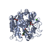
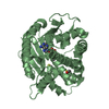
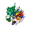
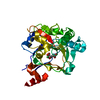
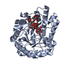
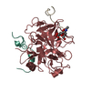
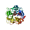
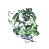
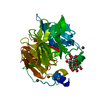
 STREPTOMYCES HYGROSCOPICUS SUBSP. ASCOMYCETICUS (bacteria)
 X-RAY DIFFRACTION /
  Authors
 Citation
 Structure visualization
 Downloads & links
 Other downloads
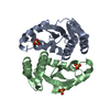
 PDBj
PDBj
 Assembly
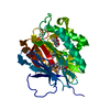
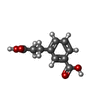
 Mass: 194.184 Da / Num. of mol.: 1 / Source method: obtained synthetically / Formula: C10H10O4
 Mass: 18.015 Da / Num. of mol.: 611 / Source method: isolated from a natural source / Formula: H2O
 Sample preparation
 / Beamline: X06SA / Wavelength: 0.97794
  Processing