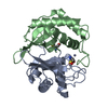
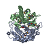
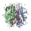
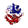
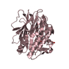
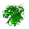
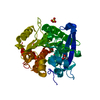
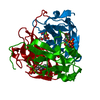
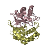
A: AUTOINDUCER-2 PRODUCTION PROTEIN B: AUTOINDUCER-2 PRODUCTION PROTEIN C: AUTOINDUCER-2 PRODUCTION PROTEIN D: AUTOINDUCER-2 PRODUCTION PROTEIN hetero molecules
Mass: 18.015 Da / Num. of mol.: 134 / Source method: isolated from a natural source / Formula: H2O
-
Experimental details
-
Experiment
Experiment
Method: X-RAY DIFFRACTION / Number of used crystals: 1
-
Sample preparation
Crystal
Density Matthews: 2.25 Å3/Da / Density % sol: 45.3 %
Crystal grow
Temperature: 295 K / Method: vapor diffusion, hanging drop / pH: 8.5 Details: Method: vapor diffusion-Hanging drop. Well Solution: PEG 4000 25%, MAGNESIUM CHLORIDE 0.2M, TRIS BUFFER 0.1M pH8.5. Drop composition: 15ul OF WELL SOLUTION, 15 ul OF PROTEIN (15mg/ml). ...Details: Method: vapor diffusion-Hanging drop. Well Solution: PEG 4000 25%, MAGNESIUM CHLORIDE 0.2M, TRIS BUFFER 0.1M pH8.5. Drop composition: 15ul OF WELL SOLUTION, 15 ul OF PROTEIN (15mg/ml). Soaking medium: PEG 4000 (27.5%), magnesium chloride 0.2M, Tris buffer 0.1M pH 8.5, Mercury Acetate 0.5mM. Soaking time: 3.5 hrs. Cryo Protectant: Lithium Formate 15%. , VAPOR DIFFUSION, HANGING DROP, temperature 295K
Resolution: 2.3→2.44 Å / Redundancy: 3.7 % / Mean I/σ(I) obs: 1.68 / Rsym value: 0.499 / % possible all: 100
-
Processing
Software
Name
Classification
SHELXS
phasing
MLPHARE
phasing
DM
modelbuilding
SHELXL-97
refinement
X-GEN
datareduction
X-GEN
datascaling
DM
phasing
Refinement
Method to determine structure: SAD(HG) / Resolution: 2.4→10 Å / Num. parameters: 18902 / Num. restraintsaints: 19055 / Cross valid method: free R throughout. / σ(F): 0 / σ(I): 0 / Stereochemistry target values: Engh & Huber Details: TWINNING REFINEMENT CARRIED OUT FIRST WITH CNS, THEN WITH SHELXL-97. FINAL TWINNING FRACTION: 0.349. THE PARTIALLY REFINED DE NOVO MODEL WAS VERIFIED AGAINST 1J6W WHEN THE COORDINATES WERE RELEASED.
In the structure databanks used in Yorodumi, some data are registered as the other names, "COVID-19 virus" and "2019-nCoV". Here are the details of the virus and the list of structure data.
Jan 31, 2019. EMDB accession codes are about to change! (news from PDBe EMDB page)
EMDB accession codes are about to change! (news from PDBe EMDB page)
The allocation of 4 digits for EMDB accession codes will soon come to an end. Whilst these codes will remain in use, new EMDB accession codes will include an additional digit and will expand incrementally as the available range of codes is exhausted. The current 4-digit format prefixed with “EMD-” (i.e. EMD-XXXX) will advance to a 5-digit format (i.e. EMD-XXXXX), and so on. It is currently estimated that the 4-digit codes will be depleted around Spring 2019, at which point the 5-digit format will come into force.
The EM Navigator/Yorodumi systems omit the EMD- prefix.
Related info.:Q: What is EMD? / ID/Accession-code notation in Yorodumi/EM Navigator
Yorodumi is a browser for structure data from EMDB, PDB, SASBDB, etc.
This page is also the successor to EM Navigator detail page, and also detail information page/front-end page for Omokage search.
The word "yorodu" (or yorozu) is an old Japanese word meaning "ten thousand". "mi" (miru) is to see.
Related info.:EMDB / PDB / SASBDB / Comparison of 3 databanks / Yorodumi Search / Aug 31, 2016. New EM Navigator & Yorodumi / Yorodumi Papers / Jmol/JSmol / Function and homology information / Changes in new EM Navigator and Yorodumi
 Movie
Movie Controller

 Yorodumi
 Open data
 Basic information
 Components
 Keywords
 Function and homology information
 Haemophilus influenzae Rd (bacteria)
 X-RAY DIFFRACTION /
  Authors
 Citation
 Structure visualization
 Downloads & links
 Other downloads
 PDBj
PDBj


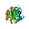
 Assembly

 Mass: 200.590 Da / Num. of mol.: 4 / Source method: obtained synthetically / Formula: Hg
 Mass: 65.409 Da / Num. of mol.: 4 / Source method: obtained synthetically / Formula: Zn
 Mass: 18.015 Da / Num. of mol.: 134 / Source method: isolated from a natural source / Formula: H2O
 Sample preparation
 / Beamline: 17-ID / Wavelength: 1 Å
 Processing