 Movie
Movie Controller

[English] 日本語
 Yorodumi
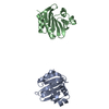
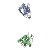
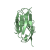
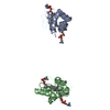
- PDB-1j4w: COMPLEX OF THE KH3 and KH4 DOMAINS OF FBP WITH A SINGLE_STRANDED ... -
+ Open data

- Basic information
| Entry | Database: PDB / ID: 1j4w | ||||||
|---|---|---|---|---|---|---|---|
| Title | COMPLEX OF THE KH3 and KH4 DOMAINS OF FBP WITH A SINGLE_STRANDED 29mer DNA OLIGONUCLEOTIDE FROM THE FUSE ELEMENT OF THE C-MYC ONCOGENE | ||||||
 Components Components |
| ||||||
 Keywords Keywords | TRANSCRIPTION/DNA / SINGLE-STRANDED DNA BINDING PROTEIN / TRANSCRIPTION FACTOR / FBP / FUSE ELEMENT / C-MYC ONCOGENE / TRANSCRIPTION-DNA COMPLEX | ||||||
| Function / homology |  Function and homology information Function and homology informationsingle-stranded DNA binding / mRNA binding / positive regulation of gene expression / regulation of transcription by RNA polymerase II / RNA binding / nucleoplasm / nucleus / cytoplasm Similarity search - Function | ||||||
| Biological species |  Homo sapiens (human) Homo sapiens (human) | ||||||
| Method | SOLUTION NMR / simulated annealing | ||||||
 Authors Authors | Clore, G.M. / Braddock, D.T. | ||||||
 Citation Citation | Journal: Nature / Year: 2002 Title: Structure and dynamics of KH domains from FBP bound to single-stranded DNA. Authors: Braddock, D.T. / Louis, J.M. / Baber, J.L. / Levens, D. / Clore, G.M. | ||||||
| History |
|
- Structure visualization
| Structure viewer | Molecule: MolmilJmol/JSmol |
|---|
- Downloads & links
-Download
| PDBx/mmCIF format | 1j4w.cif.gz | 75 KB | Display | PDBx/mmCIF format |
|---|---|---|---|---|
| PDB format | pdb1j4w.ent.gz | 55.5 KB | Display | PDB format |
| PDBx/mmJSON format | 1j4w.json.gz | Tree view | PDBx/mmJSON format | |
| Others |  Other downloads Other downloads |
-Validation report
| Summary document | 1j4w_validation.pdf.gz | 254.3 KB | Display | wwPDB validaton report |
|---|---|---|---|---|
| Full document | 1j4w_full_validation.pdf.gz | 254 KB | Display | |
| Data in XML | 1j4w_validation.xml.gz | 5.1 KB | Display | |
| Data in CIF | 1j4w_validation.cif.gz | 6.6 KB | Display | |
| Arichive directory | https://data.pdbj.org/pub/pdb/validation_reports/j4/1j4wftp://data.pdbj.org/pub/pdb/validation_reports/j4/1j4w | HTTPS FTP |
-Related structure data
| Similar structure data |
|---|


-Links
 PDBj
PDBj


- Assembly
| Deposited unit | 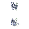
| |||||||||
|---|---|---|---|---|---|---|---|---|---|---|
| 1 |
| |||||||||
| NMR ensembles |
|
-Components
| #1: DNA chain | Mass: 8877.711 Da / Num. of mol.: 1 / Source method: obtained synthetically / Details: FROM THE FUSE ELEMENT OF THE C-MYC ONCOGENE |
|---|---|
| #2: Protein | Mass: 18638.188 Da / Num. of mol.: 1 Fragment: RESIDUES 278-447, NUMBERERED 5-174. KH3 AND KH4 DOMAINS. Mutation: C59A Source method: isolated from a genetically manipulated source Source: (gene. exp.) Homo sapiens (human) / Plasmid: PET15B / Production host:  |
-Experimental details
-Experiment
| Experiment | Method: SOLUTION NMR | ||||||||||||||||
|---|---|---|---|---|---|---|---|---|---|---|---|---|---|---|---|---|---|
| NMR experiment |
|
- Sample preparation
| Sample conditions | Ionic strength: 50 mM SODIUM PHOSPHATE / pH: 6.8 / Temperature: 308 K |
|---|---|
| Crystal grow | *PLUS Method: other / Details: NMR |
-NMR measurement
| Radiation | Protocol: SINGLE WAVELENGTH / Monochromatic (M) / Laue (L): M | ||||||||||||||||||||
|---|---|---|---|---|---|---|---|---|---|---|---|---|---|---|---|---|---|---|---|---|---|
| Radiation wavelength | Relative weight: 1 | ||||||||||||||||||||
| NMR spectrometer |
|
- Processing
| NMR software |
| ||||||||||||
|---|---|---|---|---|---|---|---|---|---|---|---|---|---|
| Refinement | Method: simulated annealing / Software ordinal: 1 Details: THE STRUCTURES WERE CALCULATED BY SIMULATED ANNEALING IN TORSION ANGLE SPACE (SCHWIETERS AND CLORE (2001) J MAGN RESON 152, 288-302) AGAINST A TARGET FUNCTION COMPRISING THE EXPERIMENTAL NMR ...Details: THE STRUCTURES WERE CALCULATED BY SIMULATED ANNEALING IN TORSION ANGLE SPACE (SCHWIETERS AND CLORE (2001) J MAGN RESON 152, 288-302) AGAINST A TARGET FUNCTION COMPRISING THE EXPERIMENTAL NMR RESTRAINTS (NOE-DERIVED INTERPROTON DISTANCE, TORSION ANGLE, 3J COUPLING, 13CALPHA/13CBETA SHIFTS AND DIPOLAR COUPLINGS). THE NON-BONDED CONTACTS IN THE TARGET FUNCTION ARE REPRESENTED BY A QUARTIC VAN DER WAALS REPULSION TERM, SUPPLEMENTED BY TORSION ANGLE (KUSZEWSKI ET AL. J. MAGN. RESON 125, 171-177 (1997)) BASE-BASE POSITIONAL (KUSZEWSKI ET AL. J AM CHEM SOC 123, 3903-3918 (2001)) DATABASE POTENTIALS OF MEAN FORCE. IN THIS ENTRY THE LAST NUMERICAL COLUMN IS THE RMS OF THE 80 INDIVIDUAL SIMULATED ANNEALING STRUCTURES (FOR EACH HALF OF THE COMPLEX) ABOUT THE MEAN COORDINATE POSITIONS: RESIDUES 75-103 OF THE PROTEIN ARE DISORDERED IN THE COMPLEX. ALTHOUGH THE SINGLE-STRANDED DNA IS B-LIKE, THE COORDINATES OF THOSE PORTIONS OF THE SS-DNA NOT IN CONTACT WITH THE PROTEIN COULD NOT BE ACCURATELY DETERMINED (BASES 201-203, 212-215 AND 223-229). THEREFORE THE COORDINATES ARE PRESENTED IN TWO HALVES: THE KH3 HALF OF THE COMPLEX (RESIDUES 1-74 OF THE PROTEIN AND BASES 216-222 OF THE SS-DNA) AND THE KH4 HALF OF THE COMPLEX (RESIDUES 104-174 OF THE PROTEIN AND BASES 204-211 OF THE SS-DNA). THE COORDINATE ACCURACY IS CALCULATED FOR THE TWO HALVES OF THE COMPLEX SEPARATELY. THE APPROXIMATE ORIENTATION OF AND SEPARATION BETWEEN THE TWO DOMAINS COULD BE DERIVED FROM ANALYSIS OF HETERONUCLEAR RELAXATION MEASUREMENTS. THE ORIENTATIONS OF THE TWO HALVES OF THE COMPLEX IN THESE COORDINATES REFLECTS THE RESULTS OF THE RELAXATION MEASUREMENTS. THE AVERAGE ORIENTATION OF THE TWO HALVES OF THE COMPLEX IS PARALLEL WITH AN AVERAGE INTERHELICAL ANGLE OF ABOUT 1 DEGREE BETWEEN THE THIRD HELIX OF EACH DOMAIN. THE OVERALL ROTATIONAL CORRELATION TIME OF THE COMPLEX IS 21.5 NS WITH A DIFFUSION ANISOTROPY OF 1.85. THE TIME SCALE FOR THE INTERDOMAIN MOTIONS IS AROUND 4 NS AND THE TWO DOMAINS WOBBLE INDEPENDENTLY IN CONES WITH SEMI-ANGLES OF ABOUT 30 DEGREES. THE OVERALL LENGTH OF THE COMPLEX IS ABOUT 100 ANGSTROMS AND THE SEPARATION BETWEEN THE TWO HALVES OF THE COMPLEX IS AROUND 35 ANGSTROMS. THE RESTRAINED REGULARIZED MEAN STRUCTURE FOR THE TWO HALVES OF THE COMPLEX IS OBTAINED BY RESTRAINED REGULARIZATION OF THE AVERAGE COORDINATES AGAINST THE SAME TARGET FUNCTION USED TO CALCULATE THE SIMULATED ANNEALING STRUCTURES. SOLVED BY MULTI HETERONUCLEAR NMR AND IS BASED ON 3153 EXPERIMENTAL NMR RESTRAINTS KH3 HALF KH4 HALF DISTANCES 1095 949 TORSION ANGLES 244 261 3JHNA COUPLINGS 33 36 13CA/CB SHIFTS 120 121 1DNH DIPOLARS 61 61 1DNC' DIPOLARS 47 39 2DHNC' DIPOLARS 46 40 BREAKDOWN OF INTRAMOLECULAR PROTEIN DISTANCE RESTRAINTS INTRARESIDUE 157 169 SEQUENTIAL 274 216 MEDIUM RANGE 247 155 LONG RANGE 260 216 BACKBONE H-BONDS 33 36 INTRA-DNA DISTANCES 41 53 INTERMOLECULAR DISTANCES 50 68 | ||||||||||||
| NMR ensemble | Conformer selection criteria: REGULARIZED MEAN STRUCTURE / Conformers calculated total number: 80 / Conformers submitted total number: 1 |
 X-PLOR NIH