 Movie
Movie Controller

[English] 日本語
 Yorodumi
- PDB-1b0w: Structural comparison of amyloidogenic light chain dimer in two c... -
+ Open data

- Basic information
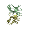
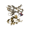
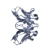
| Entry | Database: PDB / ID: 1b0w | ||||||
|---|---|---|---|---|---|---|---|
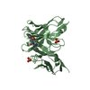
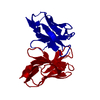
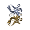
| Title | Structural comparison of amyloidogenic light chain dimer in two crystal forms with nonamyloidogenic counterparts | ||||||
 Components Components | BENCE-JONES KAPPA I PROTEIN BRE | ||||||
 Keywords Keywords | IMMUNE SYSTEM / IMMUNOGLOBULIN / AMYLOID | ||||||
| Function / homology |  Function and homology information Function and homology informationCD22 mediated BCR regulation / Fc epsilon receptor (FCERI) signaling / Classical antibody-mediated complement activation / Initial triggering of complement / FCGR activation / Role of LAT2/NTAL/LAB on calcium mobilization / immunoglobulin complex / Role of phospholipids in phagocytosis / Scavenging of heme from plasma / antigen binding ...CD22 mediated BCR regulation / Fc epsilon receptor (FCERI) signaling / Classical antibody-mediated complement activation / Initial triggering of complement / FCGR activation / Role of LAT2/NTAL/LAB on calcium mobilization / immunoglobulin complex / Role of phospholipids in phagocytosis / Scavenging of heme from plasma / antigen binding / FCERI mediated Ca+2 mobilization / FCGR3A-mediated IL10 synthesis / Regulation of Complement cascade / Antigen activates B Cell Receptor (BCR) leading to generation of second messengers / Cell surface interactions at the vascular wall / FCGR3A-mediated phagocytosis / FCERI mediated MAPK activation / Regulation of actin dynamics for phagocytic cup formation / Immunoregulatory interactions between a Lymphoid and a non-Lymphoid cell / FCERI mediated NF-kB activation / blood microparticle / Potential therapeutics for SARS / adaptive immune response / immune response / extracellular region / identical protein binding / plasma membrane Similarity search - Function | ||||||
| Biological species |  Homo sapiens (human) Homo sapiens (human) | ||||||
| Method |  X-RAY DIFFRACTION / MOLECULAR REPLACEMENT / Resolution: 1.8 Å X-RAY DIFFRACTION / MOLECULAR REPLACEMENT / Resolution: 1.8 Å | ||||||
 Authors Authors | Schormann, N. / Benson, M.D. | ||||||
 Citation Citation | Journal: Amyloid / Year: 1998 Title: Tertiary structures of amyloidogenic and non-amyloidogenic transthyretin variants: new model for amyloid fibril formation Authors: Schormann, N. / Murrell, J.R. / Benson, M.D. #1: Journal: Proc.Natl.Acad.Sci.USA / Year: 1995Title: Tertiary Structure of an Amyloid Immunoglobulin Light Chain Protein: A Proposed Model for Amyloid Fibril Formation Authors: Schormann, N. / Murrell, J.R. / Liepnieks, J.J. / Benson, M.D. | ||||||
| History |
|
- Structure visualization
| Structure viewer | Molecule: MolmilJmol/JSmol |
|---|
- Downloads & links
-Download
| PDBx/mmCIF format | 1b0w.cif.gz | 75.7 KB | Display | PDBx/mmCIF format |
|---|---|---|---|---|
| PDB format | pdb1b0w.ent.gz | 57.9 KB | Display | PDB format |
| PDBx/mmJSON format | 1b0w.json.gz | Tree view | PDBx/mmJSON format | |
| Others |  Other downloads Other downloads |
-Validation report
| Arichive directory | https://data.pdbj.org/pub/pdb/validation_reports/b0/1b0wftp://data.pdbj.org/pub/pdb/validation_reports/b0/1b0w | HTTPS FTP |
|---|
-Related structure data
| Related structure data |  1bzdC  1bzeC 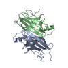 1tshC  2trhC  2tryC  1breS S: Starting model for refinement C: citing same article ( |
|---|---|
| Similar structure data |


-Links
 PDBj
PDBj


- Assembly
| Deposited unit | 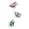
| ||||||||||||||||
|---|---|---|---|---|---|---|---|---|---|---|---|---|---|---|---|---|---|
| 1 | 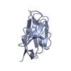
| ||||||||||||||||
| 2 | 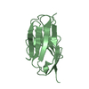
| ||||||||||||||||
| 3 | 
| ||||||||||||||||
| Unit cell |
| ||||||||||||||||
| Noncrystallographic symmetry (NCS) | NCS oper:
| ||||||||||||||||
| Details | monomer |
-Components
| #1: Antibody | Mass: 11945.124 Da / Num. of mol.: 3 / Fragment: VARIABLE DOMAIN OF LIGHT CHAIN Source method: isolated from a genetically manipulated source Source: (gene. exp.) Homo sapiens (human) / Description: PATIENT BRE / Plasmid: PCZ11Gene (production host): CDNA (GENBANK ACCESSION CODE U31344) Production host:  #2: Water | ChemComp-HOH / |  Mass: 18.015 Da / Num. of mol.: 104 / Source method: isolated from a natural source / Formula: H2O Mass: 18.015 Da / Num. of mol.: 104 / Source method: isolated from a natural source / Formula: H2OHas protein modification | Y | |
|---|
-Experimental details
-Experiment
| Experiment | Method: X-RAY DIFFRACTION / Number of used crystals: 1 |
|---|
- Sample preparation
| Crystal | Density Matthews: 3.2 Å3/Da / Density % sol: 61.15 % |
|---|---|
| Crystal grow | Method: vapor diffusion, hanging drop / pH: 6.5 Details: CRYSTALS OF BRE (20MG/ML IN 0.1M TRIS BUFFER, PH 7) WERE GROWN AT RT IN HANGING-DROPS USING 2.5M AMMONIUM SULFATE IN 0.1M CITRATE BUFFER (PH 5.5)., pH 6.5, VAPOR DIFFUSION, HANGING DROP |
-Data collection
| Diffraction | Mean temperature: 287 K |
|---|---|
| Diffraction source | Source: ROTATING ANODE / Type: RIGAKU RU200 / Wavelength: 1.5418 |
| Detector | Type: RIGAKU / Detector: IMAGE PLATE / Date: Feb 1, 1994 / Details: COLLIMATOR |
| Radiation | Monochromator: GRAPHITE / Protocol: SINGLE WAVELENGTH / Monochromatic (M) / Laue (L): M / Scattering type: x-ray |
| Radiation wavelength | Wavelength: 1.5418 Å / Relative weight: 1 |
| Reflection | Resolution: 1.75→52.5 Å / Num. obs: 35142 / % possible obs: 76 % / Observed criterion σ(I): 1 / Redundancy: 2.3 % / Biso Wilson estimate: 17.7 Å2 / Rmerge(I) obs: 0.071 / Rsym value: 0.047 / Net I/σ(I): 7.5 |
| Reflection shell | Resolution: 1.75→2 Å / Redundancy: 1.8 % / Rmerge(I) obs: 0.218 / Mean I/σ(I) obs: 1.92 / Rsym value: 0.255 / % possible all: 56 |
- Processing
| Software |
| ||||||||||||||||||||||||||||||||||||||||||||||||||||||||||||
|---|---|---|---|---|---|---|---|---|---|---|---|---|---|---|---|---|---|---|---|---|---|---|---|---|---|---|---|---|---|---|---|---|---|---|---|---|---|---|---|---|---|---|---|---|---|---|---|---|---|---|---|---|---|---|---|---|---|---|---|---|---|
| Refinement | Method to determine structure: MOLECULAR REPLACEMENT Starting model: PDB ENTRY 1BRE Resolution: 1.8→5 Å / Cross valid method: THROUGHOUT / σ(F): 2
| ||||||||||||||||||||||||||||||||||||||||||||||||||||||||||||
| Displacement parameters | Biso mean: 25.3 Å2 | ||||||||||||||||||||||||||||||||||||||||||||||||||||||||||||
| Refine analyze | Luzzati coordinate error obs: 0.28 Å / Luzzati d res low obs: 5 Å / Luzzati sigma a obs: 0.33 Å | ||||||||||||||||||||||||||||||||||||||||||||||||||||||||||||
| Refinement step | Cycle: LAST / Resolution: 1.8→5 Å
| ||||||||||||||||||||||||||||||||||||||||||||||||||||||||||||
| Refine LS restraints |
| ||||||||||||||||||||||||||||||||||||||||||||||||||||||||||||
| LS refinement shell | Resolution: 1.8→1.88 Å / Total num. of bins used: 8
| ||||||||||||||||||||||||||||||||||||||||||||||||||||||||||||
| Xplor file |
|
