[English] 日本語
Yorodumi
- PDB-2w0l: CRYSTAL STRUCTURE OF THE MUTANT H8P FROM THE RECOMBINANT VARIABLE... -

+
Open data


ID or keywords:

Loading...

-
Basic information

Entry
Database: PDB / ID: 2w0l
TitleCRYSTAL STRUCTURE OF THE MUTANT H8P FROM THE RECOMBINANT VARIABLE DOMAIN 6JAL2
ComponentsV1-22 protein
KeywordsIMMUNE SYSTEM / FIBRILS / GERM LINE / ANTIBODIES / FIBRINOGENIC
Function / homology
Function and homology information


immune response / extracellular space
Similarity search - Function
Immunoglobulin V-Type / Immunoglobulin V-set domain / Immunoglobulin V-set domain / Immunoglobulin subtype / Immunoglobulin / Ig-like domain profile. / Immunoglobulin-like domain / Immunoglobulin-like domain superfamily / Immunoglobulins / Immunoglobulin-like fold ...Immunoglobulin V-Type / Immunoglobulin V-set domain / Immunoglobulin V-set domain / Immunoglobulin subtype / Immunoglobulin / Ig-like domain profile. / Immunoglobulin-like domain / Immunoglobulin-like domain superfamily / Immunoglobulins / Immunoglobulin-like fold / Immunoglobulin-like / Sandwich / Mainly Beta
Similarity search - Domain/homology
Biological speciesHomo sapiens (human)
MethodX-RAY DIFFRACTION / SYNCHROTRON / MOLECULAR REPLACEMENT / Resolution: 2.2 Å
AuthorsRudino-Pinera, E. / Gonzalezrubio-Garrido, P. / Horjales, E.
CitationJournal: To be Published
Title: Crystal Structure of the Variable Domain 6Ajl2
Authors: Hdz-Santollo, A. / Rudino-Pinera, E. / Fuentes-Silva, D. / Gonzalezrubio-Garrido, P. / Delpozoyauner, L. / Becerril, B. / Horjales, E. / Rodriguez-Romero, A.
History
DepositionAug 19, 2008Deposition site: PDBE / Processing site: PDBE
Revision 1.0Nov 17, 2009Provider: repository / Type: Initial release
Revision 1.1May 8, 2011Group: Version format compliance
Revision 1.2Jul 13, 2011Group: Version format compliance
Revision 1.3Nov 7, 2018Group: Data collection / Database references ...Data collection / Database references / Source and taxonomy / Structure summary
Category: entity / entity_name_com ...entity / entity_name_com / entity_src_gen / entity_src_nat / struct_ref / struct_ref_seq / struct_ref_seq_dif
Item: _entity.pdbx_description / _entity.src_method
Revision 1.4Dec 13, 2023Group: Data collection / Database references ...Data collection / Database references / Other / Refinement description
Category: chem_comp_atom / chem_comp_bond ...chem_comp_atom / chem_comp_bond / database_2 / pdbx_database_status / pdbx_initial_refinement_model / struct_ncs_dom_lim
Item: _database_2.pdbx_DOI / _database_2.pdbx_database_accession ..._database_2.pdbx_DOI / _database_2.pdbx_database_accession / _pdbx_database_status.status_code_sf / _struct_ncs_dom_lim.beg_auth_comp_id / _struct_ncs_dom_lim.beg_label_asym_id / _struct_ncs_dom_lim.beg_label_comp_id / _struct_ncs_dom_lim.beg_label_seq_id / _struct_ncs_dom_lim.end_auth_comp_id / _struct_ncs_dom_lim.end_label_asym_id / _struct_ncs_dom_lim.end_label_comp_id / _struct_ncs_dom_lim.end_label_seq_id
Remark 700 
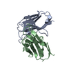
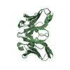
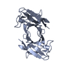
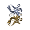
SHEET THE SHEET STRUCTURE OF THIS MOLECULE IS BIFURCATED. IN ORDER TO REPRESENT THIS FEATURE IN ... SHEET THE SHEET STRUCTURE OF THIS MOLECULE IS BIFURCATED. IN ORDER TO REPRESENT THIS FEATURE IN THE SHEET RECORDS BELOW, TWO SHEETS ARE DEFINED.

-
Structure visualization

Structure viewerMolecule:
MolmilJmol/JSmol

Downloads & links

-
Assembly

Deposited unit
A: V1-22 protein
B: V1-22 protein
C: V1-22 protein
D: V1-22 protein


Theoretical massNumber of molelcules
Total (without water)47,6844
Polymers47,6844
Non-polymers00
Water5,693316
1
A: V1-22 protein
C: V1-22 protein


Theoretical massNumber of molelcules
Total (without water)23,8422
Polymers23,8422
Non-polymers00
Water362
TypeNameSymmetry operationNumber
identity operation1_555x,y,z1
Buried area1180 Å2
ΔGint-6.94 kcal/mol
Surface area10410 Å2
MethodPISA
2
B: V1-22 protein
D: V1-22 protein


Theoretical massNumber of molelcules
Total (without water)23,8422
Polymers23,8422
Non-polymers00
Water362
TypeNameSymmetry operationNumber
identity operation1_555x,y,z1
Buried area1080 Å2
ΔGint-6.54 kcal/mol
Surface area10500 Å2
MethodPISA
Unit cell
Length a, b, c (Å)40.940, 85.495, 69.152
Angle α, β, γ (deg.)90.00, 91.20, 90.00
Int Tables number4
Space group name H-MP1211
Noncrystallographic symmetry (NCS)NCS domain:
IDEns-IDDetails
11A
21B
31C
41D

NCS domain segments:

Component-ID: 1 / Ens-ID: 1 / Beg auth comp-ID: ASN / Beg label comp-ID: ASN / End auth comp-ID: LEU / End label comp-ID: LEU / Refine code: 1 / Auth seq-ID: 1 - 107 / Label seq-ID: 1 - 111

Dom-IDAuth asym-IDLabel asym-ID
1AA
2BB
3CC
4DD

-
Components

#1: Antibody
V1-22 protein


Mass: 11920.878 Da / Num. of mol.: 4 / Mutation: YES
Source method: isolated from a genetically manipulated source
Source: (gene. exp.) Homo sapiens (human) / Gene: V1-22 / Production host: Escherichia coli (E. coli) / References: UniProt: Q5NV88
#2: Water ChemComp-HOH / water


Mass: 18.015 Da / Num. of mol.: 316 / Source method: isolated from a natural source / Formula: H2O
Compound detailsENGINEERED RESIDUE IN CHAIN A, HIS 8 TO PRO ENGINEERED RESIDUE IN CHAIN B, HIS 8 TO PRO ENGINEERED ...ENGINEERED RESIDUE IN CHAIN A, HIS 8 TO PRO ENGINEERED RESIDUE IN CHAIN B, HIS 8 TO PRO ENGINEERED RESIDUE IN CHAIN C, HIS 8 TO PRO ENGINEERED RESIDUE IN CHAIN D, HIS 8 TO PRO
Sequence details
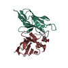
THIS ENTRY COMPRISE THE FIRST 98 RESIDUES OF UNIPROT ENTRY PLUS MUTATION H8P

-
Experimental details

-
Experiment

ExperimentMethod: X-RAY DIFFRACTION / Number of used crystals: 1

-
Sample preparation

CrystalDensity Matthews: 2.84 Å3/Da / Density % sol: 56.64 % / Description: NONE

-
Data collection

DiffractionMean temperature: 100 K
Diffraction sourceSource: SYNCHROTRON / Site: NSLS / Beamline: X6A / Wavelength: 0.9795
DetectorType: ADSC CCD / Detector: CCD / Date: May 24, 2005
RadiationProtocol: SINGLE WAVELENGTH / Monochromatic (M) / Laue (L): M / Scattering type: x-ray
Radiation wavelengthWavelength: 0.9795 Å / Relative weight: 1
ReflectionResolution: 2.2→53.7 Å / Num. obs: 22584 / % possible obs: 78.8 % / Observed criterion σ(I): 0 / Redundancy: 1.6 % / Rmerge(I) obs: 0.04 / Net I/σ(I): 16.1
Reflection shellResolution: 2.2→2.32 Å / Redundancy: 1.5 % / Rmerge(I) obs: 0.1 / Mean I/σ(I) obs: 6.6 / % possible all: 43.9

-
Processing

Software
NameVersionClassification
REFMAC5.2.0019refinement
MOSFLMdata reduction
SCALAdata scaling
MOLREPphasing
RefinementMethod to determine structure: MOLECULAR REPLACEMENT
Starting model: PDB ENTRY 2CD0
Resolution: 2.2→35.56 Å / Cor.coef. Fo:Fc: 0.926 / Cor.coef. Fo:Fc free: 0.891 / SU B: 6.408 / SU ML: 0.164 / Cross valid method: THROUGHOUT / ESU R: 0.421 / ESU R Free: 0.25 / Stereochemistry target values: MAXIMUM LIKELIHOOD / Details: HYDROGENS HAVE BEEN ADDED IN THE RIDING POSITIONS.
RfactorNum. reflection% reflectionSelection details
Rfree0.245 1056 5.1 %RANDOM
Rwork0.206 ---
obs0.208 19558 85.1 %-
Solvent computationIon probe radii: 0.8 Å / Shrinkage radii: 0.8 Å / VDW probe radii: 1.2 Å / Solvent model: MASK
Displacement parametersBiso mean: 20.53 Å2
Baniso -1Baniso -2Baniso -3
1-0.01 Å20 Å20.02 Å2
2---0.01 Å20 Å2
3---0 Å2
Refinement stepCycle: LAST / Resolution: 2.2→35.56 Å
ProteinNucleic acidLigandSolventTotal
Num. atoms3348 0 0 316 3664
Refine LS restraints
Refine-IDTypeDev idealDev ideal targetNumber
X-RAY DIFFRACTIONr_bond_refined_d0.020.0223420
X-RAY DIFFRACTIONr_bond_other_d
X-RAY DIFFRACTIONr_angle_refined_deg2.0541.9554656
X-RAY DIFFRACTIONr_angle_other_deg
X-RAY DIFFRACTIONr_dihedral_angle_1_deg12.0565436
X-RAY DIFFRACTIONr_dihedral_angle_2_deg41.11824.722144
X-RAY DIFFRACTIONr_dihedral_angle_3_deg16.25215524
X-RAY DIFFRACTIONr_dihedral_angle_4_deg14.2791516
X-RAY DIFFRACTIONr_chiral_restr0.1810.2520
X-RAY DIFFRACTIONr_gen_planes_refined0.0090.022628
X-RAY DIFFRACTIONr_gen_planes_other
X-RAY DIFFRACTIONr_nbd_refined0.2850.21276
X-RAY DIFFRACTIONr_nbd_other
X-RAY DIFFRACTIONr_nbtor_refined0.30.22272
X-RAY DIFFRACTIONr_nbtor_other
X-RAY DIFFRACTIONr_xyhbond_nbd_refined0.4210.2280
X-RAY DIFFRACTIONr_xyhbond_nbd_other
X-RAY DIFFRACTIONr_metal_ion_refined
X-RAY DIFFRACTIONr_metal_ion_other
X-RAY DIFFRACTIONr_symmetry_vdw_refined0.3470.229
X-RAY DIFFRACTIONr_symmetry_vdw_other
X-RAY DIFFRACTIONr_symmetry_hbond_refined0.1650.210
X-RAY DIFFRACTIONr_symmetry_hbond_other
X-RAY DIFFRACTIONr_symmetry_metal_ion_refined
X-RAY DIFFRACTIONr_symmetry_metal_ion_other
X-RAY DIFFRACTIONr_mcbond_it1.1011.52268
X-RAY DIFFRACTIONr_mcbond_other
X-RAY DIFFRACTIONr_mcangle_it1.85523592
X-RAY DIFFRACTIONr_mcangle_other
X-RAY DIFFRACTIONr_scbond_it2.13231327
X-RAY DIFFRACTIONr_scbond_other
X-RAY DIFFRACTIONr_scangle_it3.14.51064
X-RAY DIFFRACTIONr_scangle_other
X-RAY DIFFRACTIONr_long_range_B_refined
X-RAY DIFFRACTIONr_long_range_B_other
X-RAY DIFFRACTIONr_rigid_bond_restr
X-RAY DIFFRACTIONr_sphericity_free
X-RAY DIFFRACTIONr_sphericity_bonded
Refine LS restraints NCS

Ens-ID: 1 / Number: 837 / Refine-ID: X-RAY DIFFRACTION

Dom-IDAuth asym-IDTypeRms dev position (Å)Weight position
1Atight positional0.140.05
2Btight positional0.080.05
3Ctight positional0.090.05
4Dtight positional0.090.05
1Atight thermal0.190.5
2Btight thermal0.180.5
3Ctight thermal0.180.5
4Dtight thermal0.190.5
LS refinement shellResolution: 2.2→2.26 Å / Total num. of bins used: 20 /
RfactorNum. reflection
Rfree0.363 33
Rwork0.23 707

+
About Yorodumi

-
News

-
Feb 9, 2022. New format data for meta-information of EMDB entries

New format data for meta-information of EMDB entries

  • Version 3 of the EMDB header file is now the official format.
  • The previous official version 1.9 will be removed from the archive.

Related info.:EMDB header

External links:wwPDB to switch to version 3 of the EMDB data model

-
Aug 12, 2020. Covid-19 info

Covid-19 info

URL: https://pdbjlvh1.pdbj.org/emnavi/covid19.php

New page: Covid-19 featured information page in EM Navigator.

Related info.:Covid-19 info / Mar 5, 2020. Novel coronavirus structure data

+
Mar 5, 2020. Novel coronavirus structure data

Novel coronavirus structure data

Related info.:Yorodumi Speices / Aug 12, 2020. Covid-19 info

External links:COVID-19 featured content - PDBj / Molecule of the Month (242):Coronavirus Proteases

+
Jan 31, 2019. EMDB accession codes are about to change! (news from PDBe EMDB page)

EMDB accession codes are about to change! (news from PDBe EMDB page)

  • The allocation of 4 digits for EMDB accession codes will soon come to an end. Whilst these codes will remain in use, new EMDB accession codes will include an additional digit and will expand incrementally as the available range of codes is exhausted. The current 4-digit format prefixed with “EMD-” (i.e. EMD-XXXX) will advance to a 5-digit format (i.e. EMD-XXXXX), and so on. It is currently estimated that the 4-digit codes will be depleted around Spring 2019, at which point the 5-digit format will come into force.
  • The EM Navigator/Yorodumi systems omit the EMD- prefix.

Related info.:Q: What is EMD? / ID/Accession-code notation in Yorodumi/EM Navigator

External links:EMDB Accession Codes are Changing Soon! / Contact to PDBj

+
Jul 12, 2017. Major update of PDB

Major update of PDB

  • wwPDB released updated PDB data conforming to the new PDBx/mmCIF dictionary.
  • This is a major update changing the version number from 4 to 5, and with Remediation, in which all the entries are updated.
  • In this update, many items about electron microscopy experimental information are reorganized (e.g. em_software).
  • Now, EM Navigator and Yorodumi are based on the updated data.

External links:wwPDB Remediation / Enriched Model Files Conforming to OneDep Data Standards Now Available in the PDB FTP Archive

-
Yorodumi

Thousand views of thousand structures

  • Yorodumi is a browser for structure data from EMDB, PDB, SASBDB, etc.
  • This page is also the successor to EM Navigator detail page, and also detail information page/front-end page for Omokage search.
  • The word "yorodu" (or yorozu) is an old Japanese word meaning "ten thousand". "mi" (miru) is to see.

Related info.:EMDB / PDB / SASBDB / Comparison of 3 databanks / Yorodumi Search / Aug 31, 2016. New EM Navigator & Yorodumi / Yorodumi Papers / Jmol/JSmol / Function and homology information / Changes in new EM Navigator and Yorodumi

Read more