 Movie
Movie Controller

[English] 日本語
 Yorodumi
- PDB-1j35: Crystal Structure of Ca(II)-bound Gla Domain of Factor IX Complex... -
+ Open data

- Basic information
| Entry | Database: PDB / ID: 1j35 | ||||||
|---|---|---|---|---|---|---|---|
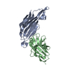
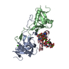
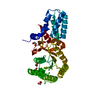
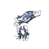
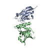
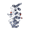
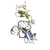
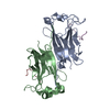
| Title | Crystal Structure of Ca(II)-bound Gla Domain of Factor IX Complexed with Binding Protein | ||||||
 Components Components |
| ||||||
 Keywords Keywords | Protein binding/Blood clotting / MAGNESIUM ION / CALCIUM ION / GLA DOMAIN / Protein binding-Blood clotting COMPLEX | ||||||
| Function / homology |  Function and homology information Function and homology informationcoagulation factor IXa / zymogen activation / blood coagulation / toxin activity / endopeptidase activity / endoplasmic reticulum lumen / serine-type endopeptidase activity / calcium ion binding / magnesium ion binding / proteolysis ...coagulation factor IXa / zymogen activation / blood coagulation / toxin activity / endopeptidase activity / endoplasmic reticulum lumen / serine-type endopeptidase activity / calcium ion binding / magnesium ion binding / proteolysis / : / extracellular region / metal ion binding Similarity search - Function | ||||||
| Biological species |  Trimeresurus flavoviridis (habu) Trimeresurus flavoviridis (habu) | ||||||
| Method |  X-RAY DIFFRACTION / SYNCHROTRON / MOLECULAR REPLACEMENT / Resolution: 1.8 Å X-RAY DIFFRACTION / SYNCHROTRON / MOLECULAR REPLACEMENT / Resolution: 1.8 Å | ||||||
 Authors Authors | Shikamoto, Y. / Morita, T. / Fujimoto, Z. / Mizuno, H. | ||||||
 Citation Citation | Journal: J.Biol.Chem. / Year: 2003 Title: Crystal Structure of Mg2+- and Ca2+-bound Gla Domain of Factor IX Complexed with Binding Protein Authors: Shikamoto, Y. / Morita, T. / Fujimoto, Z. / Mizuno, H. #1: Journal: Proc.Natl.Acad.Sci.USA / Year: 2001Title: Crystal structure of an anticoagulant protein in complex with the Gla domain of factor X Authors: Mizuno, H. / Fujimoto, Z. / Atoda, H. / Morita, T. #2: Journal: J.Mol.Biol. / Year: 1999Title: Crystal structure of coagulation factor IX-binding protein from habu snake venom at 2.6 A: implication of central loop swapping based on deletion in the linker region Authors: Mizuno, H. / Fujimoto, Z. / Koizumi, M. / Kano, H. / Atoda, H. / Morita, T. | ||||||
| History |
|
- Structure visualization
| Structure viewer | Molecule: MolmilJmol/JSmol |
|---|
- Downloads & links
-Download
| PDBx/mmCIF format | 1j35.cif.gz | 87.8 KB | Display | PDBx/mmCIF format |
|---|---|---|---|---|
| PDB format | pdb1j35.ent.gz | 64 KB | Display | PDB format |
| PDBx/mmJSON format | 1j35.json.gz | Tree view | PDBx/mmJSON format | |
| Others |  Other downloads Other downloads |
-Validation report
| Arichive directory | https://data.pdbj.org/pub/pdb/validation_reports/j3/1j35ftp://data.pdbj.org/pub/pdb/validation_reports/j3/1j35 | HTTPS FTP |
|---|
-Related structure data
| Related structure data |  1j34C 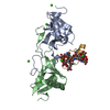 1iodS S: Starting model for refinement C: citing same article ( |
|---|---|
| Similar structure data |


-Links
 PDBj
PDBj


- Assembly
| Deposited unit | 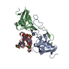
| ||||||||
|---|---|---|---|---|---|---|---|---|---|
| 1 |
| ||||||||
| Unit cell |
|
-Components
| #1: Protein | Mass: 14655.184 Da / Num. of mol.: 1 / Source method: isolated from a natural source / Source: (natural) Trimeresurus flavoviridis (habu) / Secretion: venom / References: UniProt: P23806, UniProt: Q7LZ71*PLUS | ||
|---|---|---|---|
| #2: Protein | Mass: 14455.071 Da / Num. of mol.: 1 / Source method: isolated from a natural source / Source: (natural) Trimeresurus flavoviridis (habu) / Secretion: venom / References: UniProt: P23807 | ||
| #3: Protein/peptide | Mass: 6177.313 Da / Num. of mol.: 1 / Fragment: GLA DOMAIN / Source method: isolated from a natural source / Source: (natural) | ||
| #4: Chemical | ChemComp-CA /   Mass: 40.078 Da / Num. of mol.: 10 / Source method: obtained synthetically / Formula: Ca Mass: 40.078 Da / Num. of mol.: 10 / Source method: obtained synthetically / Formula: Ca#5: Water | ChemComp-HOH / |  Mass: 18.015 Da / Num. of mol.: 266 / Source method: isolated from a natural source / Formula: H2O Mass: 18.015 Da / Num. of mol.: 266 / Source method: isolated from a natural source / Formula: H2O |
-Experimental details
-Experiment
| Experiment | Method: X-RAY DIFFRACTION / Number of used crystals: 1 |
|---|
- Sample preparation
| Crystal | Density Matthews: 2.04 Å3/Da / Density % sol: 39.3 % | ||||||||||||||||||||||||||||||||||||||||||
|---|---|---|---|---|---|---|---|---|---|---|---|---|---|---|---|---|---|---|---|---|---|---|---|---|---|---|---|---|---|---|---|---|---|---|---|---|---|---|---|---|---|---|---|
| Crystal grow | Temperature: 293 K / Method: microbatch / pH: 8 Details: PEG 6000, Tris-HCl, calcium chloride, pH 8, MICROBATCH, temperature 293K | ||||||||||||||||||||||||||||||||||||||||||
| Crystal grow | *PLUS Temperature: 20 ℃ / pH: 8 / Method: batch method | ||||||||||||||||||||||||||||||||||||||||||
| Components of the solutions | *PLUS
|
-Data collection
| Diffraction | Mean temperature: 100 K |
|---|---|
| Diffraction source | Source: SYNCHROTRON / Site: SPring-8  / Beamline: BL41XU / Wavelength: 0.72 Å / Beamline: BL41XU / Wavelength: 0.72 Å |
| Detector | Type: MARRESEARCH / Detector: CCD / Date: May 30, 2001 |
| Radiation | Protocol: SINGLE WAVELENGTH / Monochromatic (M) / Laue (L): M / Scattering type: x-ray |
| Radiation wavelength | Wavelength: 0.72 Å / Relative weight: 1 |
| Reflection | Resolution: 1.8→20 Å / Num. all: 27532 / Num. obs: 27532 / % possible obs: 93.9 % / Observed criterion σ(I): 0 / Redundancy: 9.5 % / Biso Wilson estimate: 18.6 Å2 / Rmerge(I) obs: 0.046 / Net I/σ(I): 13.7 |
| Reflection shell | Resolution: 1.8→1.88 Å / Rmerge(I) obs: 0.252 / Num. unique all: 2639 / % possible all: 72.4 |
| Reflection | *PLUS % possible obs: 94 % |
- Processing
| Software |
| ||||||||||||||||||||||||||||||||||||
|---|---|---|---|---|---|---|---|---|---|---|---|---|---|---|---|---|---|---|---|---|---|---|---|---|---|---|---|---|---|---|---|---|---|---|---|---|---|
| Refinement | Method to determine structure: MOLECULAR REPLACEMENT Starting model: 1IOD Resolution: 1.8→19.54 Å / Rfactor Rfree error: 0.005 / Data cutoff high absF: 445935.96 / Data cutoff low absF: 0 / Isotropic thermal model: RESTRAINED / Cross valid method: THROUGHOUT / σ(F): 0 / Stereochemistry target values: Engh & Huber
| ||||||||||||||||||||||||||||||||||||
| Solvent computation | Solvent model: FLAT MODEL / Bsol: 50.8158 Å2 / ksol: 0.321533 e/Å3 | ||||||||||||||||||||||||||||||||||||
| Displacement parameters | Biso mean: 32.6 Å2
| ||||||||||||||||||||||||||||||||||||
| Refine analyze |
| ||||||||||||||||||||||||||||||||||||
| Refinement step | Cycle: LAST / Resolution: 1.8→19.54 Å
| ||||||||||||||||||||||||||||||||||||
| Refine LS restraints |
| ||||||||||||||||||||||||||||||||||||
| LS refinement shell | Resolution: 1.8→1.91 Å / Rfactor Rfree error: 0.017 / Total num. of bins used: 6
| ||||||||||||||||||||||||||||||||||||
| Xplor file |
| ||||||||||||||||||||||||||||||||||||
| Refinement | *PLUS Lowest resolution: 20 Å / Rfactor Rfree: 0.27 | ||||||||||||||||||||||||||||||||||||
| Solvent computation | *PLUS | ||||||||||||||||||||||||||||||||||||
| Displacement parameters | *PLUS | ||||||||||||||||||||||||||||||||||||
| Refine LS restraints | *PLUS
|
