 Movie
Movie Controller

[English] 日本語
 Yorodumi
- PDB-1iw0: Crystal structure of a heme oxygenase (HmuO) from Corynebacterium... -
+ Open data

- Basic information
| Entry | Database: PDB / ID: 1iw0 | |||||||||
|---|---|---|---|---|---|---|---|---|---|---|
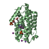
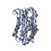
| Title | Crystal structure of a heme oxygenase (HmuO) from Corynebacterium diphtheriae complexed with heme in the ferric state | |||||||||
 Components Components | Heme oxygenase | |||||||||
 Keywords Keywords | OXIDOREDUCTASE / alpha helix / bacterial iron acquisition / RIKEN Structural Genomics/Proteomics Initiative / RSGI / Structural Genomics | |||||||||
| Function / homology |  Function and homology information Function and homology informationheme oxygenase (biliverdin-producing) / heme oxidation / heme oxygenase (decyclizing) activity / heme catabolic process / response to oxidative stress / heme binding / metal ion binding Similarity search - Function | |||||||||
| Biological species |  Corynebacterium diphtheriae (bacteria) Corynebacterium diphtheriae (bacteria) | |||||||||
| Method |  X-RAY DIFFRACTION / SYNCHROTRON / Resolution: 1.4 Å X-RAY DIFFRACTION / SYNCHROTRON / Resolution: 1.4 Å | |||||||||
 Authors Authors | Hirotsu, S. / Unno, M. / Chu, G.C. / Lee, D.S. / Park, S.Y. / Shiro, Y. / Ikeda-Saito, M. / RIKEN Structural Genomics/Proteomics Initiative (RSGI) | |||||||||
 Citation Citation | Journal: J.Biol.Chem. / Year: 2004 Title: The crystal structures of the ferric and ferrous forms of the heme complex of HmuO, a heme oxygenase of Corynebacterium diphtheriae. Authors: Hirotsu, S. / Chu, G.C. / Unno, M. / Lee, D.S. / Yoshida, T. / Park, S.Y. / Shiro, Y. / Ikeda-Saito, M. | |||||||||
| History |
|
- Structure visualization
| Structure viewer | Molecule: MolmilJmol/JSmol |
|---|
- Downloads & links
-Download
| PDBx/mmCIF format | 1iw0.cif.gz | 284.1 KB | Display | PDBx/mmCIF format |
|---|---|---|---|---|
| PDB format | pdb1iw0.ent.gz | 229.3 KB | Display | PDB format |
| PDBx/mmJSON format | 1iw0.json.gz | Tree view | PDBx/mmJSON format | |
| Others |  Other downloads Other downloads |
-Validation report
| Arichive directory | https://data.pdbj.org/pub/pdb/validation_reports/iw/1iw0ftp://data.pdbj.org/pub/pdb/validation_reports/iw/1iw0 | HTTPS FTP |
|---|
-Related structure data


-Links
 PDBj
PDBj- Assembly
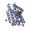
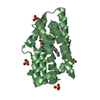
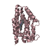
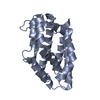
| Deposited unit | 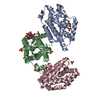
| ||||||||
|---|---|---|---|---|---|---|---|---|---|
| 1 | 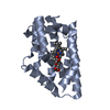
| ||||||||
| 2 | 
| ||||||||
| 3 | 
| ||||||||
| Unit cell |
| ||||||||
| Details | Biological assembly is a monomer |
-Components
| #1: Protein | Mass: 24170.158 Da / Num. of mol.: 3 Source method: isolated from a genetically manipulated source Source: (gene. exp.) Corynebacterium diphtheriae (bacteria) / Plasmid: pMW172 / Species (production host): Escherichia coli / Production host: References: UniProt: P71119, heme oxygenase (biliverdin-producing) #2: Polysaccharide | beta-D-fructofuranose-(2-1)-alpha-D-glucopyranose / sucrose |   Source method: isolated from a genetically manipulated source Details: oligosaccharide with reducing-end-to-reducing-end glycosidic bond References: sucrose #3: Chemical |   Mass: 616.487 Da / Num. of mol.: 3 / Source method: obtained synthetically / Formula: C34H32FeN4O4 Mass: 616.487 Da / Num. of mol.: 3 / Source method: obtained synthetically / Formula: C34H32FeN4O4#4: Chemical | ChemComp-SO4 /   Mass: 96.063 Da / Num. of mol.: 6 / Source method: obtained synthetically / Formula: SO4 Mass: 96.063 Da / Num. of mol.: 6 / Source method: obtained synthetically / Formula: SO4#5: Water | ChemComp-HOH / |  Mass: 18.015 Da / Num. of mol.: 655 / Source method: isolated from a natural source / Formula: H2O Mass: 18.015 Da / Num. of mol.: 655 / Source method: isolated from a natural source / Formula: H2O |
|---|
-Experimental details
-Experiment
| Experiment | Method: X-RAY DIFFRACTION / Number of used crystals: 1 |
|---|
- Sample preparation
| Crystal | Density Matthews: 2.18 Å3/Da / Density % sol: 43.05 % | ||||||||||||||||||||||||||||||||||||
|---|---|---|---|---|---|---|---|---|---|---|---|---|---|---|---|---|---|---|---|---|---|---|---|---|---|---|---|---|---|---|---|---|---|---|---|---|---|
| Crystal grow | Temperature: 303 K / Method: vapor diffusion, sitting drop / pH: 5.8 Details: ammonium sulfate, NaI, MES, pH 5.8, VAPOR DIFFUSION, SITTING DROP, temperature 303K | ||||||||||||||||||||||||||||||||||||
| Crystal grow | *PLUS pH: 7 | ||||||||||||||||||||||||||||||||||||
| Components of the solutions | *PLUS
|
-Data collection
| Diffraction | Mean temperature: 98 K |
|---|---|
| Diffraction source | Source: SYNCHROTRON / Site: SPring-8  / Beamline: BL45PX / Wavelength: 0.9 Å / Beamline: BL45PX / Wavelength: 0.9 Å |
| Detector | Type: RIGAKU JUPITER 210 / Detector: CCD / Date: Dec 6, 2001 / Details: mirrors |
| Radiation | Monochromator: Diamond / Protocol: SINGLE WAVELENGTH / Monochromatic (M) / Laue (L): M / Scattering type: x-ray |
| Radiation wavelength | Wavelength: 0.9 Å / Relative weight: 1 |
| Reflection | Resolution: 1.3→99 Å / Num. all: 261943 / Num. obs: 146822 / % possible obs: 85 % / Rmerge(I) obs: 0.034 / Net I/σ(I): 18.08 |
| Reflection shell | Resolution: 1.3→1.34 Å / Rmerge(I) obs: 0.297 / Mean I/σ(I) obs: 0.87 / % possible all: 58.6 |
| Reflection | *PLUS Lowest resolution: 99 Å / Num. measured all: 542325 |
| Reflection shell | *PLUS % possible obs: 58.6 % |
- Processing
| Software |
| ||||||||||||||||||||||||||||||||||||||||||||||||||||||||||||||||||||||||||||||||||||||||||||||||||||||||||||||||||||||||||||||||||
|---|---|---|---|---|---|---|---|---|---|---|---|---|---|---|---|---|---|---|---|---|---|---|---|---|---|---|---|---|---|---|---|---|---|---|---|---|---|---|---|---|---|---|---|---|---|---|---|---|---|---|---|---|---|---|---|---|---|---|---|---|---|---|---|---|---|---|---|---|---|---|---|---|---|---|---|---|---|---|---|---|---|---|---|---|---|---|---|---|---|---|---|---|---|---|---|---|---|---|---|---|---|---|---|---|---|---|---|---|---|---|---|---|---|---|---|---|---|---|---|---|---|---|---|---|---|---|---|---|---|---|---|
| Refinement | Resolution: 1.4→12 Å / Cor.coef. Fo:Fc: 0.965 / Cor.coef. Fo:Fc free: 0.957 / SU B: 3.213 / SU ML: 0.064 / Cross valid method: THROUGHOUT / ESU R: 0.078 / ESU R Free: 0.066 / Stereochemistry target values: MAXIMUM LIKELIHOOD / Details: HYDROGENS HAVE BEEN ADDED IN THE RIDING POSITIONS
| ||||||||||||||||||||||||||||||||||||||||||||||||||||||||||||||||||||||||||||||||||||||||||||||||||||||||||||||||||||||||||||||||||
| Solvent computation | Ion probe radii: 0.8 Å / Shrinkage radii: 0.8 Å / VDW probe radii: 1.4 Å / Solvent model: BABINET MODEL WITH MASK | ||||||||||||||||||||||||||||||||||||||||||||||||||||||||||||||||||||||||||||||||||||||||||||||||||||||||||||||||||||||||||||||||||
| Displacement parameters | Biso mean: 15.799 Å2
| ||||||||||||||||||||||||||||||||||||||||||||||||||||||||||||||||||||||||||||||||||||||||||||||||||||||||||||||||||||||||||||||||||
| Refinement step | Cycle: LAST / Resolution: 1.4→12 Å
| ||||||||||||||||||||||||||||||||||||||||||||||||||||||||||||||||||||||||||||||||||||||||||||||||||||||||||||||||||||||||||||||||||
| Refine LS restraints |
| ||||||||||||||||||||||||||||||||||||||||||||||||||||||||||||||||||||||||||||||||||||||||||||||||||||||||||||||||||||||||||||||||||
| LS refinement shell | Resolution: 1.4→1.436 Å / Total num. of bins used: 20 /
| ||||||||||||||||||||||||||||||||||||||||||||||||||||||||||||||||||||||||||||||||||||||||||||||||||||||||||||||||||||||||||||||||||
| Refinement | *PLUS Highest resolution: 1.4 Å / Lowest resolution: 12 Å / Rfactor Rfree: 0.192 / Rfactor Rwork: 0.165 | ||||||||||||||||||||||||||||||||||||||||||||||||||||||||||||||||||||||||||||||||||||||||||||||||||||||||||||||||||||||||||||||||||
| Solvent computation | *PLUS | ||||||||||||||||||||||||||||||||||||||||||||||||||||||||||||||||||||||||||||||||||||||||||||||||||||||||||||||||||||||||||||||||||
| Displacement parameters | *PLUS | ||||||||||||||||||||||||||||||||||||||||||||||||||||||||||||||||||||||||||||||||||||||||||||||||||||||||||||||||||||||||||||||||||
| Refine LS restraints | *PLUS
|
