 Movie
Movie Controller


+ Open data

- Basic information
| Entry | Database: PDB / ID: 1is6 | |||||||||
|---|---|---|---|---|---|---|---|---|---|---|
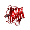
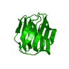
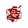
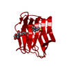
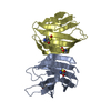
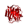
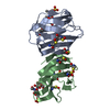
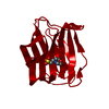
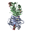
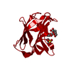
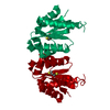
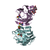
| Title | MES-Liganded Congerin II | |||||||||
 Components Components | Congerin II | |||||||||
 Keywords Keywords | SUGAR BINDING PROTEIN / MES complex / BETA SANDWICH | |||||||||
| Function / homology |  Function and homology information Function and homology informationgalactoside binding / laminin binding / carbohydrate binding / extracellular space Similarity search - Function | |||||||||
| Biological species |  Conger myriaster (whitespotted conger) Conger myriaster (whitespotted conger) | |||||||||
| Method |  X-RAY DIFFRACTION / MOLECULAR REPLACEMENT / Resolution: 1.7 Å X-RAY DIFFRACTION / MOLECULAR REPLACEMENT / Resolution: 1.7 Å | |||||||||
 Authors Authors | Shirai, T. / Matsui, Y. / Shionyu-Mitsuyama, C. / Yamane, T. / Kamiya, H. / Ishii, C. / Ogawa, T. / Muramoto, K. | |||||||||
 Citation Citation | Journal: J.MOL.BIOL. / Year: 2002 Title: Crystal structure of a conger eel galectin (congerin II) at 1.45 A resolution: Implication for the accelerated evolution of a new ligand-binding site following gene duplication Authors: Shirai, T. / Matsui, Y. / Shionyu-Mitsuyama, C. / Yamane, T. / Kamiya, H. / Ishii, C. / Ogawa, T. / Muramoto, K. | |||||||||
| History |
|
- Structure visualization
| Structure viewer | Molecule: MolmilJmol/JSmol |
|---|
- Downloads & links
-Download
| PDBx/mmCIF format | 1is6.cif.gz | 40.3 KB | Display | PDBx/mmCIF format |
|---|---|---|---|---|
| PDB format | pdb1is6.ent.gz | 27.9 KB | Display | PDB format |
| PDBx/mmJSON format | 1is6.json.gz | Tree view | PDBx/mmJSON format | |
| Others |  Other downloads Other downloads |
-Validation report
| Summary document | 1is6_validation.pdf.gz | 377.8 KB | Display | wwPDB validaton report |
|---|---|---|---|---|
| Full document | 1is6_full_validation.pdf.gz | 379.4 KB | Display | |
| Data in XML | 1is6_validation.xml.gz | 4.7 KB | Display | |
| Data in CIF | 1is6_validation.cif.gz | 6.8 KB | Display | |
| Arichive directory | https://data.pdbj.org/pub/pdb/validation_reports/is/1is6ftp://data.pdbj.org/pub/pdb/validation_reports/is/1is6 | HTTPS FTP |
-Related structure data


-Links
 PDBj
PDBj- Assembly
| Deposited unit | 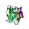
| ||||||||
|---|---|---|---|---|---|---|---|---|---|
| 1 | 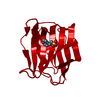
| ||||||||
| Unit cell |
| ||||||||
| Components on special symmetry positions |
| ||||||||
| Details | The second part of the biological assembly is generated by the two fold axis : y, x, -z+1. |
-Components
| #1: Protein | Mass: 15354.119 Da / Num. of mol.: 1 Source method: isolated from a genetically manipulated source Source: (gene. exp.) Conger myriaster (whitespotted conger) / Plasmid: pTV118N / Production host:  |
|---|---|
| #2: Chemical | ChemComp-MES /   Mass: 195.237 Da / Num. of mol.: 1 / Source method: obtained synthetically / Formula: C6H13NO4S / Comment: pH buffer*YM Mass: 195.237 Da / Num. of mol.: 1 / Source method: obtained synthetically / Formula: C6H13NO4S / Comment: pH buffer*YM |
| #3: Water | ChemComp-HOH /  Mass: 18.015 Da / Num. of mol.: 69 / Source method: isolated from a natural source / Formula: H2O Mass: 18.015 Da / Num. of mol.: 69 / Source method: isolated from a natural source / Formula: H2O |
-Experimental details
-Experiment
| Experiment | Method: X-RAY DIFFRACTION / Number of used crystals: 1 |
|---|
- Sample preparation
| Crystal | Density Matthews: 2.48 Å3/Da / Density % sol: 50.39 % | ||||||||||||||||||||||||||||||||||||||||||
|---|---|---|---|---|---|---|---|---|---|---|---|---|---|---|---|---|---|---|---|---|---|---|---|---|---|---|---|---|---|---|---|---|---|---|---|---|---|---|---|---|---|---|---|
| Crystal grow | Temperature: 291 K / Method: vapor diffusion, hanging drop / pH: 6.5 Details: magnesium sulfate, MES, pH 6.5, VAPOR DIFFUSION, HANGING DROP, temperature 291K | ||||||||||||||||||||||||||||||||||||||||||
| Crystal grow | *PLUS Temperature: 18 ℃ | ||||||||||||||||||||||||||||||||||||||||||
| Components of the solutions | *PLUS
|
-Data collection
| Diffraction | Mean temperature: 291 K |
|---|---|
| Diffraction source | Source: ROTATING ANODE / Type: RIGAKU RU300 / Wavelength: 1.54 Å |
| Detector | Type: RIGAKU RAXIS IV / Detector: IMAGE PLATE / Date: Dec 25, 2000 |
| Radiation | Protocol: SINGLE WAVELENGTH / Monochromatic (M) / Laue (L): M / Scattering type: x-ray |
| Radiation wavelength | Wavelength: 1.54 Å / Relative weight: 1 |
| Reflection | Resolution: 1.7→99 Å / Num. all: 17071 / Num. obs: 17071 / % possible obs: 96.4 % / Observed criterion σ(F): 0 / Observed criterion σ(I): 0 / Rmerge(I) obs: 0.047 / Net I/σ(I): 30.38 |
| Reflection shell | Resolution: 1.7→1.76 Å / Rmerge(I) obs: 0.194 / Mean I/σ(I) obs: 4.9 / Num. unique all: 1414 / % possible all: 82 |
| Reflection | *PLUS Rmerge(I) obs: 0.047 |
| Reflection shell | *PLUS % possible obs: 82 % / Num. unique obs: 1414 / Rmerge(I) obs: 0.194 |
- Processing
| Software |
| |||||||||||||||||||||||||
|---|---|---|---|---|---|---|---|---|---|---|---|---|---|---|---|---|---|---|---|---|---|---|---|---|---|---|
| Refinement | Method to determine structure: MOLECULAR REPLACEMENT Starting model: Congerin II Lactose and Mes complex Resolution: 1.7→8 Å / σ(F): 3 / Stereochemistry target values: Engh & Huber
| |||||||||||||||||||||||||
| Refinement step | Cycle: LAST / Resolution: 1.7→8 Å
| |||||||||||||||||||||||||
| Refine LS restraints |
| |||||||||||||||||||||||||
| LS refinement shell | Resolution: 1.7→1.76 Å
| |||||||||||||||||||||||||
| Refinement | *PLUS Highest resolution: 1.7 Å / Lowest resolution: 8 Å / Rfactor all: 0.192 / Rfactor obs: 0.188 / Rfactor Rfree: 0.232 / Rfactor Rwork: 0.187 | |||||||||||||||||||||||||
| Solvent computation | *PLUS | |||||||||||||||||||||||||
| Displacement parameters | *PLUS | |||||||||||||||||||||||||
| Refine LS restraints | *PLUS
| |||||||||||||||||||||||||
| LS refinement shell | *PLUS Rfactor Rfree: 0.234 / Rfactor Rwork: 0.237 |
