 Movie
Movie Controller

[English] 日本語
 Yorodumi
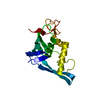
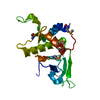
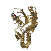
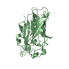
- PDB-1iqv: Crystal Structure Analysis of the archaebacterial ribosomal protein S7 -
+ Open data

- Basic information
| Entry | Database: PDB / ID: 1iqv | ||||||
|---|---|---|---|---|---|---|---|
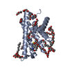
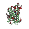
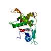
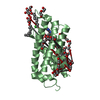
| Title | Crystal Structure Analysis of the archaebacterial ribosomal protein S7 | ||||||
 Components Components | RIBOSOMAL PROTEIN S7 | ||||||
 Keywords Keywords | RIBOSOME / RIBOSOMAL PROTEIN / RNA-BINDING / DECODING CENTER / HELIX-TURN-HELIX | ||||||
| Function / homology |  Function and homology information Function and homology informationsmall ribosomal subunit / rRNA binding / structural constituent of ribosome / translation Similarity search - Function | ||||||
| Biological species |   Pyrococcus horikoshii (archaea) Pyrococcus horikoshii (archaea) | ||||||
| Method |  X-RAY DIFFRACTION / SYNCHROTRON / MOLECULAR REPLACEMENT / Resolution: 2.1 Å X-RAY DIFFRACTION / SYNCHROTRON / MOLECULAR REPLACEMENT / Resolution: 2.1 Å | ||||||
 Authors Authors | Hosaka, H. / Yao, M. / Kimura, M. / Tanaka, I. | ||||||
 Citation Citation | Journal: J.Biochem. / Year: 2001 Title: The structure of the archaebacterial ribosomal protein S7 and its possible interaction with 16S rRNA. Authors: Hosaka, H. / Yao, M. / Kimura, M. / Tanaka, I. | ||||||
| History |
|
- Structure visualization
| Structure viewer | Molecule: MolmilJmol/JSmol |
|---|
- Downloads & links
-Download
| PDBx/mmCIF format | 1iqv.cif.gz | 48.8 KB | Display | PDBx/mmCIF format |
|---|---|---|---|---|
| PDB format | pdb1iqv.ent.gz | 33.7 KB | Display | PDB format |
| PDBx/mmJSON format | 1iqv.json.gz | Tree view | PDBx/mmJSON format | |
| Others |  Other downloads Other downloads |
-Validation report
| Summary document | 1iqv_validation.pdf.gz | 367.9 KB | Display | wwPDB validaton report |
|---|---|---|---|---|
| Full document | 1iqv_full_validation.pdf.gz | 370.5 KB | Display | |
| Data in XML | 1iqv_validation.xml.gz | 4.9 KB | Display | |
| Data in CIF | 1iqv_validation.cif.gz | 7.5 KB | Display | |
| Arichive directory | https://data.pdbj.org/pub/pdb/validation_reports/iq/1iqvftp://data.pdbj.org/pub/pdb/validation_reports/iq/1iqv | HTTPS FTP |
-Related structure data
| Related structure data | 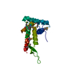 1husS S: Starting model for refinement |
|---|---|
| Similar structure data |


-Links
 PDBj
PDBj

- Assembly
| Deposited unit | 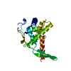
| ||||||||
|---|---|---|---|---|---|---|---|---|---|
| 1 |
| ||||||||
| Unit cell |
|
-Components
| #1: Protein | Mass: 25029.232 Da / Num. of mol.: 1 Source method: isolated from a genetically manipulated source Source: (gene. exp.) Pyrococcus horikoshii (archaea) / Gene: PH1541 / Plasmid: pET22b / Production host:  |
|---|---|
| #2: Water | ChemComp-HOH /  Mass: 18.015 Da / Num. of mol.: 131 / Source method: isolated from a natural source / Formula: H2O Mass: 18.015 Da / Num. of mol.: 131 / Source method: isolated from a natural source / Formula: H2O |
-Experimental details
-Experiment
| Experiment | Method: X-RAY DIFFRACTION / Number of used crystals: 1 |
|---|
- Sample preparation
| Crystal | Density Matthews: 2.1 Å3/Da / Density % sol: 41.9 % | ||||||||||||||||||||||||||||||||||||||||||
|---|---|---|---|---|---|---|---|---|---|---|---|---|---|---|---|---|---|---|---|---|---|---|---|---|---|---|---|---|---|---|---|---|---|---|---|---|---|---|---|---|---|---|---|
| Crystal grow | Temperature: 293 K / Method: vapor diffusion, hanging drop / pH: 7 Details: PEG3000, sodium chloride, pH 7.0, VAPOR DIFFUSION, HANGING DROP, temperature 293.0K | ||||||||||||||||||||||||||||||||||||||||||
| Crystal grow | *PLUS | ||||||||||||||||||||||||||||||||||||||||||
| Components of the solutions | *PLUS
|
-Data collection
| Diffraction | Mean temperature: 100 K |
|---|---|
| Diffraction source | Source: SYNCHROTRON / Site: SPring-8  / Beamline: BL44XU / Wavelength: 0.9 Å / Beamline: BL44XU / Wavelength: 0.9 Å |
| Detector | Type: OXFORD PX210 / Detector: CCD / Date: Mar 14, 2001 |
| Radiation | Monochromator: Monochromator + Mirror / Protocol: SINGLE WAVELENGTH / Monochromatic (M) / Laue (L): M / Scattering type: x-ray |
| Radiation wavelength | Wavelength: 0.9 Å / Relative weight: 1 |
| Reflection | Resolution: 2.1→31 Å / Num. all: 14407 / Num. obs: 14407 / % possible obs: 99.8 % / Observed criterion σ(F): 0 / Observed criterion σ(I): 0 / Redundancy: 9.7 % / Biso Wilson estimate: 41.4 Å2 / Rmerge(I) obs: 0.081 / Rsym value: 0.077 / Net I/σ(I): 7.2 |
| Reflection shell | Resolution: 2.1→2.21 Å / Redundancy: 7.1 % / Rmerge(I) obs: 0.262 / Mean I/σ(I) obs: 2.9 / Num. unique all: 2048 / Rsym value: 0.243 / % possible all: 99.8 |
| Reflection | *PLUS Lowest resolution: 40 Å / Num. measured all: 139697 |
| Reflection shell | *PLUS % possible obs: 99.8 % |
- Processing
| Software |
| |||||||||||||||||||||||||||
|---|---|---|---|---|---|---|---|---|---|---|---|---|---|---|---|---|---|---|---|---|---|---|---|---|---|---|---|---|
| Refinement | Method to determine structure: MOLECULAR REPLACEMENT Starting model: PDB ENTRY 1HUS Resolution: 2.1→10 Å / Isotropic thermal model: isotropic / Cross valid method: THROUGHOUT / σ(F): 2 / Stereochemistry target values: Engh & Huber
| |||||||||||||||||||||||||||
| Solvent computation | Bsol: 66.8 Å2 / ksol: 0.436 e/Å3 | |||||||||||||||||||||||||||
| Displacement parameters | Biso mean: 35.7 Å2
| |||||||||||||||||||||||||||
| Refine analyze |
| |||||||||||||||||||||||||||
| Refinement step | Cycle: LAST / Resolution: 2.1→10 Å
| |||||||||||||||||||||||||||
| Refine LS restraints |
| |||||||||||||||||||||||||||
| LS refinement shell | Resolution: 2.1→2.17 Å / Total num. of bins used: 10
| |||||||||||||||||||||||||||
| Xplor file | Serial no: 1 / Param file: protein_rep.param / Topol file: protein.top | |||||||||||||||||||||||||||
| Software | *PLUS Name: CNS / Classification: refinement | |||||||||||||||||||||||||||
| Refinement | *PLUS Highest resolution: 2.1 Å / Lowest resolution: 10 Å / Num. reflection obs: 14240 / σ(F): 2 / % reflection Rfree: 10 % / Rfactor obs: 0.196 / Rfactor Rfree: 0.23 | |||||||||||||||||||||||||||
| Solvent computation | *PLUS | |||||||||||||||||||||||||||
| Displacement parameters | *PLUS Biso mean: 35.7 Å2 | |||||||||||||||||||||||||||
| Refine LS restraints | *PLUS
| |||||||||||||||||||||||||||
| LS refinement shell | *PLUS Rfactor Rfree: 0.265 / % reflection Rfree: 10 % / Rfactor Rwork: 0.243 |
