 Movie
Movie Controller


+ Open data

- Basic information
| Entry | Database: PDB / ID: 1ijh | ||||||
|---|---|---|---|---|---|---|---|
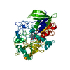
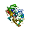
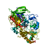
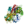
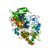
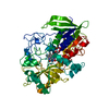
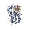
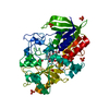
| Title | CHOLESTEROL OXIDASE FROM STREPTOMYCES ASN485LEU MUTANT | ||||||
 Components Components | CHOLESTEROL OXIDASE | ||||||
 Keywords Keywords | OXIDOREDUCTASE / FLAVOENZYME / STEROID METABOLISM | ||||||
| Function / homology |  Function and homology information Function and homology informationcholesterol oxidase / cholesterol oxidase activity / steroid Delta-isomerase / steroid Delta-isomerase activity / cholesterol catabolic process / flavin adenine dinucleotide binding / extracellular region Similarity search - Function | ||||||
| Biological species |  Streptomyces sp. (bacteria) Streptomyces sp. (bacteria) | ||||||
| Method |  X-RAY DIFFRACTION / OTHER / Resolution: 1.53 Å X-RAY DIFFRACTION / OTHER / Resolution: 1.53 Å | ||||||
 Authors Authors | Vrielink, A. / Lario, P.I. | ||||||
 Citation Citation | Journal: Biochemistry / Year: 2001 Title: The presence of a hydrogen bond between asparagine 485 and the pi system of FAD modulates the redox potential in the reaction catalyzed by cholesterol oxidase. Authors: Yin, Y. / Sampson, N.S. / Vrielink, A. / Lario, P.I. | ||||||
| History |
|
- Structure visualization
| Structure viewer | Molecule: MolmilJmol/JSmol |
|---|
- Downloads & links
-Download
| PDBx/mmCIF format | 1ijh.cif.gz | 128.2 KB | Display | PDBx/mmCIF format |
|---|---|---|---|---|
| PDB format | pdb1ijh.ent.gz | 95.3 KB | Display | PDB format |
| PDBx/mmJSON format | 1ijh.json.gz | Tree view | PDBx/mmJSON format | |
| Others |  Other downloads Other downloads |
-Validation report
| Arichive directory | https://data.pdbj.org/pub/pdb/validation_reports/ij/1ijhftp://data.pdbj.org/pub/pdb/validation_reports/ij/1ijh | HTTPS FTP |
|---|
-Related structure data
| Related structure data |  1b4vS S: Starting model for refinement |
|---|---|
| Similar structure data |


-Links
 PDBj
PDBj


- Assembly
| Deposited unit | 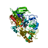
| ||||||||
|---|---|---|---|---|---|---|---|---|---|
| 1 |
| ||||||||
| Unit cell |
|
-Components
| #1: Protein | Mass: 54968.730 Da / Num. of mol.: 1 / Mutation: N485L Source method: isolated from a genetically manipulated source Details: FAD COFACTOR NON-COVALENTLY BOUND TO THE ENZYME / Source: (gene. exp.) Streptomyces sp. (bacteria) / Gene: choA / Plasmid: pCO237 / Production host: |
|---|---|
| #2: Chemical | ChemComp-FAD /   Mass: 785.550 Da / Num. of mol.: 1 / Source method: obtained synthetically / Formula: C27H33N9O15P2 / Comment: FAD*YM Mass: 785.550 Da / Num. of mol.: 1 / Source method: obtained synthetically / Formula: C27H33N9O15P2 / Comment: FAD*YM |
| #3: Water | ChemComp-HOH /  Mass: 18.015 Da / Num. of mol.: 607 / Source method: isolated from a natural source / Formula: H2O Mass: 18.015 Da / Num. of mol.: 607 / Source method: isolated from a natural source / Formula: H2O |
-Experimental details
-Experiment
| Experiment | Method: X-RAY DIFFRACTION / Number of used crystals: 1 |
|---|
- Sample preparation
| Crystal | Density Matthews: 1 Å3/Da / Density % sol: 40.5 % | ||||||||||||||||||||||||||||||||||||
|---|---|---|---|---|---|---|---|---|---|---|---|---|---|---|---|---|---|---|---|---|---|---|---|---|---|---|---|---|---|---|---|---|---|---|---|---|---|
| Crystal grow | Temperature: 290 K / Method: vapor diffusion, hanging drop / pH: 5.2 Details: PEG 8000, SODIUM CACODYLATE, MNSO4, pH 5.2, VAPOR DIFFUSION, HANGING DROP, temperature 290K | ||||||||||||||||||||||||||||||||||||
| Crystal grow | *PLUS Temperature: 17 ℃ / pH: 7 / Details: Yue, K., (1999) Biochemistry, 38, 4277. | ||||||||||||||||||||||||||||||||||||
| Components of the solutions | *PLUS
|
-Data collection
| Diffraction | Mean temperature: 86 K |
|---|---|
| Diffraction source | Source: ROTATING ANODE / Type: RIGAKU RU200 / Wavelength: 1.5418 |
| Detector | Type: MARRESEARCH / Detector: IMAGE PLATE / Date: Sep 9, 1999 / Details: MIRRORS |
| Radiation | Protocol: SINGLE WAVELENGTH / Monochromatic (M) / Laue (L): M / Scattering type: x-ray |
| Radiation wavelength | Wavelength: 1.5418 Å / Relative weight: 1 |
| Reflection | Resolution: 1.52→25 Å / Num. obs: 63695 / % possible obs: 92.8 % / Observed criterion σ(F): 0 / Observed criterion σ(I): 0 / Redundancy: 4.1 % / Biso Wilson estimate: 21.8 Å2 / Rmerge(I) obs: 0.045 / Net I/σ(I): 25.5 |
| Reflection shell | Resolution: 1.52→1.58 Å / Redundancy: 3.6 % / Rmerge(I) obs: 0.265 / Mean I/σ(I) obs: 4.6 / Num. unique all: 5048 / % possible all: 73.8 |
| Reflection | *PLUS Lowest resolution: 25 Å |
- Processing
| Software |
| |||||||||||||||||||||||||||||||||
|---|---|---|---|---|---|---|---|---|---|---|---|---|---|---|---|---|---|---|---|---|---|---|---|---|---|---|---|---|---|---|---|---|---|---|
| Refinement | Method to determine structure: OTHER Starting model: 1B4V Resolution: 1.53→25 Å / Num. parameters: 18258 / Num. restraintsaints: 16261 / Cross valid method: FREE R / σ(F): 0 / σ(I): 0 / Stereochemistry target values: ENGH AND HUBER Details: The following are residues that were not located in the experiment: ASP A 6, ASN A 7, GLY A 8, THR A 507, ALA A 508, SER A 509. Some very mobile side chains were modeled at either 50% or ...Details: The following are residues that were not located in the experiment: ASP A 6, ASN A 7, GLY A 8, THR A 507, ALA A 508, SER A 509. Some very mobile side chains were modeled at either 50% or zero occupancy. The occupancy of absent side chain atoms was set to zero for the following: RES 396 ARG NE->end has zero occupancy. RES 436 GLN CG->end has zero occupancy. Partially occupied side chain atoms modeled at 50% for the following: RES 146 ARG atoms CZ, NH1,NH2; RES 163 LYS atoms CE,NZ; RES 273 LYS atoms CG,CD,CE,NZ; RES 278 LYS atoms CG,CD,CE,NZ; RES 279 GLU atoms CG,CD,OE1,OE2; RES 396 ARG atoms CG,CD (REST ZERO); RES 404 ASP atoms CG, OD1,OD2; RES 468 LYS atoms CG,CD,CE, NZ; Regarding the close contacts in remark 500: ARG 396 NH1 (zero occupancy); GLU 279 OE2 50% occupancy; ARG 202 NE very aniostropic B = 33; All of the flagged waters are modeled at 50% occupancy.
| |||||||||||||||||||||||||||||||||
| Solvent computation | Solvent model: MOEWS & KRETSINGER, J.MOL.BIOL.91(1973)201-228 | |||||||||||||||||||||||||||||||||
| Displacement parameters | Biso mean: 16.8 Å2 | |||||||||||||||||||||||||||||||||
| Refine analyze | Num. disordered residues: 15 / Occupancy sum hydrogen: 0 / Occupancy sum non hydrogen: 4450.5 | |||||||||||||||||||||||||||||||||
| Refinement step | Cycle: LAST / Resolution: 1.53→25 Å
| |||||||||||||||||||||||||||||||||
| Refine LS restraints |
| |||||||||||||||||||||||||||||||||
| Software | *PLUS Name: SHELXL-97 / Classification: refinement | |||||||||||||||||||||||||||||||||
| Refinement | *PLUS Lowest resolution: 25 Å / Num. reflection obs: 57331 / σ(F): 0 / Num. reflection Rfree: 6632 / % reflection Rfree: 10 % / Rfactor all: 0.166 / Rfactor obs: 0.163 | |||||||||||||||||||||||||||||||||
| Solvent computation | *PLUS | |||||||||||||||||||||||||||||||||
| Displacement parameters | *PLUS | |||||||||||||||||||||||||||||||||
| Refine LS restraints | *PLUS Type: s_plane_restr / Dev ideal: 0.03 |
