 Movie
Movie Controller


+ Open data

- Basic information
| Entry | Database: PDB / ID: 1b4v | ||||||
|---|---|---|---|---|---|---|---|
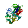
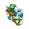
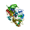
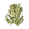
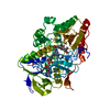
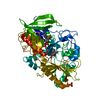
| Title | CHOLESTEROL OXIDASE FROM STREPTOMYCES | ||||||
 Components Components | PROTEIN (CHOLESTEROL OXIDASE) | ||||||
 Keywords Keywords | OXIDOREDUCTASE / FLAVOENZYME / STEROID METABOLISM | ||||||
| Function / homology |  Function and homology information Function and homology informationcholesterol oxidase / cholesterol oxidase activity / steroid Delta-isomerase / steroid Delta-isomerase activity / cholesterol catabolic process / flavin adenine dinucleotide binding / extracellular region Similarity search - Function | ||||||
| Biological species |  Streptomyces sp. (bacteria) Streptomyces sp. (bacteria) | ||||||
| Method |  X-RAY DIFFRACTION / MOLECULAR REPLACEMENT / Resolution: 1.5 Å X-RAY DIFFRACTION / MOLECULAR REPLACEMENT / Resolution: 1.5 Å | ||||||
 Authors Authors | Vrielink, A. / Yue, Q.K. | ||||||
 Citation Citation | Journal: Biochemistry / Year: 1999 Title: Crystal structure determination of cholesterol oxidase from Streptomyces and structural characterization of key active site mutants. Authors: Yue, Q.K. / Kass, I.J. / Sampson, N.S. / Vrielink, A. | ||||||
| History |
|
- Structure visualization
| Structure viewer | Molecule: MolmilJmol/JSmol |
|---|
- Downloads & links
-Download
| PDBx/mmCIF format | 1b4v.cif.gz | 127.3 KB | Display | PDBx/mmCIF format |
|---|---|---|---|---|
| PDB format | pdb1b4v.ent.gz | 94.5 KB | Display | PDB format |
| PDBx/mmJSON format | 1b4v.json.gz | Tree view | PDBx/mmJSON format | |
| Others |  Other downloads Other downloads |
-Validation report
| Arichive directory | https://data.pdbj.org/pub/pdb/validation_reports/b4/1b4vftp://data.pdbj.org/pub/pdb/validation_reports/b4/1b4v | HTTPS FTP |
|---|
-Related structure data
| Related structure data | 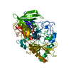 1b8sC  1cboC 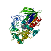 1cc2C  3coxS S: Starting model for refinement C: citing same article ( |
|---|---|
| Similar structure data |


-Links
 PDBj
PDBj


- Assembly
| Deposited unit | 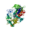
| ||||||||
|---|---|---|---|---|---|---|---|---|---|
| 1 |
| ||||||||
| Unit cell |
|
-Components
| #1: Protein | Mass: 54969.676 Da / Num. of mol.: 1 / Source method: isolated from a natural source / Details: FAD COFACTOR NON-COVALENTLY BOUND TO THE ENZYME / Source: (natural) Streptomyces sp. (bacteria) / References: UniProt: P12676, cholesterol oxidase |
|---|---|
| #2: Chemical | ChemComp-FAD /   Mass: 785.550 Da / Num. of mol.: 1 / Source method: obtained synthetically / Formula: C27H33N9O15P2 / Comment: FAD*YM Mass: 785.550 Da / Num. of mol.: 1 / Source method: obtained synthetically / Formula: C27H33N9O15P2 / Comment: FAD*YM |
| #3: Water | ChemComp-HOH /  Mass: 18.015 Da / Num. of mol.: 664 / Source method: isolated from a natural source / Formula: H2O Mass: 18.015 Da / Num. of mol.: 664 / Source method: isolated from a natural source / Formula: H2O |
| Sequence details | THE SEQUENCE OF CHOLESTEROL OXIDASE FROM STREPTOMYCES SP FOUND IN THE SEQUENCE DATA BASE IS A ...THE SEQUENCE OF CHOLESTERO |
-Experimental details
-Experiment
| Experiment | Method: X-RAY DIFFRACTION / Number of used crystals: 1 |
|---|
- Sample preparation
| Crystal | Density Matthews: 2.2 Å3/Da / Density % sol: 44 % | ||||||||||||||||||||||||||||||||||||
|---|---|---|---|---|---|---|---|---|---|---|---|---|---|---|---|---|---|---|---|---|---|---|---|---|---|---|---|---|---|---|---|---|---|---|---|---|---|
| Crystal grow | Method: vapor diffusion, hanging drop / pH: 5.2 Details: PRECIPITANT CONDITIONS: 10- 12% PEG 8000, 100MM SODIUM CACODYLATE PH 5.2, 75MM MNSO4 PROTEIN CONCENTRATION 8.5MG/ML, VAPOR DIFFUSION, HANGING DROP | ||||||||||||||||||||||||||||||||||||
| Crystal grow | *PLUS Temperature: 17 ℃ / pH: 7 | ||||||||||||||||||||||||||||||||||||
| Components of the solutions | *PLUS
|
-Data collection
| Diffraction | Mean temperature: 115 K |
|---|---|
| Diffraction source | Source: ROTATING ANODE / Type: RIGAKU RU300 / Wavelength: 1.5418 |
| Detector | Type: RIGAKU RAXIS II / Detector: IMAGE PLATE / Date: Oct 1, 1996 |
| Radiation | Monochromator: GRAPHITE CRYSTAL / Protocol: SINGLE WAVELENGTH / Monochromatic (M) / Laue (L): M / Scattering type: x-ray |
| Radiation wavelength | Wavelength: 1.5418 Å / Relative weight: 1 |
| Reflection | Resolution: 1.5→22.3 Å / Num. obs: 62747 / % possible obs: 87.4 % / Redundancy: 6.4 % / Biso Wilson estimate: 14.9 Å2 / Rmerge(I) obs: 0.045 / Net I/σ(I): 27.5 |
| Reflection shell | Resolution: 1.5→1.55 Å / Redundancy: 0.31 % / Rmerge(I) obs: 0.22 / Mean I/σ(I) obs: 2 / % possible all: 30.9 |
| Reflection | *PLUS Num. measured all: 402969 |
- Processing
| Software |
| ||||||||||||||||||||||||||||||||||||||||||||||||||||||||||||||||||||||||||||||||
|---|---|---|---|---|---|---|---|---|---|---|---|---|---|---|---|---|---|---|---|---|---|---|---|---|---|---|---|---|---|---|---|---|---|---|---|---|---|---|---|---|---|---|---|---|---|---|---|---|---|---|---|---|---|---|---|---|---|---|---|---|---|---|---|---|---|---|---|---|---|---|---|---|---|---|---|---|---|---|---|---|---|
| Refinement | Method to determine structure: MOLECULAR REPLACEMENT Starting model: 3COX Resolution: 1.5→20 Å / Rfactor Rfree error: 0.003 / Data cutoff high absF: 1000000 / Data cutoff low absF: 0.001 / Isotropic thermal model: RESTRAINED / Cross valid method: THROUGHOUT / σ(F): 0
| ||||||||||||||||||||||||||||||||||||||||||||||||||||||||||||||||||||||||||||||||
| Displacement parameters | Biso mean: 12.4 Å2 | ||||||||||||||||||||||||||||||||||||||||||||||||||||||||||||||||||||||||||||||||
| Refine analyze |
| ||||||||||||||||||||||||||||||||||||||||||||||||||||||||||||||||||||||||||||||||
| Refinement step | Cycle: LAST / Resolution: 1.5→20 Å
| ||||||||||||||||||||||||||||||||||||||||||||||||||||||||||||||||||||||||||||||||
| Refine LS restraints |
| ||||||||||||||||||||||||||||||||||||||||||||||||||||||||||||||||||||||||||||||||
| LS refinement shell | Resolution: 1.5→1.57 Å / Rfactor Rfree error: 0.023 / Total num. of bins used: 8
| ||||||||||||||||||||||||||||||||||||||||||||||||||||||||||||||||||||||||||||||||
| Xplor file |
| ||||||||||||||||||||||||||||||||||||||||||||||||||||||||||||||||||||||||||||||||
| Software | *PLUS Name: X-PLOR / Version: 3.843 / Classification: refinement | ||||||||||||||||||||||||||||||||||||||||||||||||||||||||||||||||||||||||||||||||
| Refinement | *PLUS Highest resolution: 1.5 Å / Lowest resolution: 20 Å / σ(F): 0 / % reflection Rfree: 10.1 % / Rfactor Rfree: 0.2 | ||||||||||||||||||||||||||||||||||||||||||||||||||||||||||||||||||||||||||||||||
| Solvent computation | *PLUS | ||||||||||||||||||||||||||||||||||||||||||||||||||||||||||||||||||||||||||||||||
| Displacement parameters | *PLUS Biso mean: 12.4 Å2 | ||||||||||||||||||||||||||||||||||||||||||||||||||||||||||||||||||||||||||||||||
| Refine LS restraints | *PLUS
| ||||||||||||||||||||||||||||||||||||||||||||||||||||||||||||||||||||||||||||||||
| LS refinement shell | *PLUS Highest resolution: 1.5 Å / % reflection Rfree: 10.1 % |
