 Movie
Movie Controller


+ Open data

- Basic information
| Entry | Database: PDB / ID: 1iho | ||||||
|---|---|---|---|---|---|---|---|
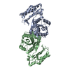
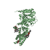
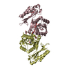
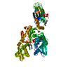
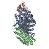
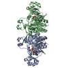
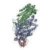
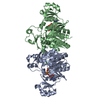
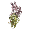
| Title | CRYSTAL APO-STRUCTURE OF PANTOTHENATE SYNTHETASE FROM E. COLI | ||||||
 Components Components | PANTOATE--BETA-ALANINE LIGASE | ||||||
 Keywords Keywords | LIGASE / Rossmann fold / dimer / apo / HIGH / KSMKS / flexible domains / multidomain | ||||||
| Function / homology |  Function and homology information Function and homology informationpantoate-beta-alanine ligase (AMP-forming) / pantoate-beta-alanine ligase activity / pantothenate biosynthetic process / protein homodimerization activity / ATP binding / identical protein binding / cytosol Similarity search - Function | ||||||
| Biological species |  | ||||||
| Method |  X-RAY DIFFRACTION / SYNCHROTRON / MAD / Resolution: 1.7 Å X-RAY DIFFRACTION / SYNCHROTRON / MAD / Resolution: 1.7 Å | ||||||
 Authors Authors | von Delft, F. / Lewendon, A. / Dhanaraj, V. / Blundell, T.L. / Abell, C. / Smith, A. | ||||||
 Citation Citation | Journal: Structure / Year: 2001 Title: The crystal structure of E. coli pantothenate synthetase confirms it as a member of the cytidylyltransferase superfamily. Authors: von Delft, F. / Lewendon, A. / Dhanaraj, V. / Blundell, T.L. / Abell, C. / Smith, A.G. | ||||||
| History |
|
- Structure visualization
| Structure viewer | Molecule: MolmilJmol/JSmol |
|---|
- Downloads & links
-Download
| PDBx/mmCIF format | 1iho.cif.gz | 134.5 KB | Display | PDBx/mmCIF format |
|---|---|---|---|---|
| PDB format | pdb1iho.ent.gz | 103.9 KB | Display | PDB format |
| PDBx/mmJSON format | 1iho.json.gz | Tree view | PDBx/mmJSON format | |
| Others |  Other downloads Other downloads |
-Validation report
| Arichive directory | https://data.pdbj.org/pub/pdb/validation_reports/ih/1ihoftp://data.pdbj.org/pub/pdb/validation_reports/ih/1iho | HTTPS FTP |
|---|
-Related structure data
| Similar structure data |
|---|

-Links
 PDBj
PDBj- Assembly
| Deposited unit | 
| ||||||||
|---|---|---|---|---|---|---|---|---|---|
| 1 |
| ||||||||
| Unit cell |
| ||||||||
| Details | The biological assembly is completely represented by the dimer in the asymmetric unit. |
-Components
| #1: Protein | Mass: 31639.689 Da / Num. of mol.: 2 Source method: isolated from a genetically manipulated source Source: (gene. exp.) References: UniProt: P31663, pantoate-beta-alanine ligase (AMP-forming) #2: Chemical | ChemComp-TRS / |   Mass: 122.143 Da / Num. of mol.: 1 / Source method: obtained synthetically / Formula: C4H12NO3 / Comment: pH buffer*YM Mass: 122.143 Da / Num. of mol.: 1 / Source method: obtained synthetically / Formula: C4H12NO3 / Comment: pH buffer*YM#3: Chemical |   Mass: 62.068 Da / Num. of mol.: 2 / Source method: obtained synthetically / Formula: C2H6O2 Mass: 62.068 Da / Num. of mol.: 2 / Source method: obtained synthetically / Formula: C2H6O2#4: Water | ChemComp-HOH / |  Mass: 18.015 Da / Num. of mol.: 614 / Source method: isolated from a natural source / Formula: H2O Mass: 18.015 Da / Num. of mol.: 614 / Source method: isolated from a natural source / Formula: H2O |
|---|
-Experimental details
-Experiment
| Experiment | Method: X-RAY DIFFRACTION / Number of used crystals: 2 |
|---|
- Sample preparation
| Crystal | Density Matthews: 2.97 Å3/Da / Density % sol: 58 % | |||||||||||||||
|---|---|---|---|---|---|---|---|---|---|---|---|---|---|---|---|---|
| Crystal grow | Temperature: 298 K / Method: vapor diffusion, hanging drop / pH: 8 Details: PEG 4000 (4-6%), Tris buffer pH 8 (50 mM), VAPOR DIFFUSION, HANGING DROP, temperature 298K | |||||||||||||||
| Crystal grow | *PLUS Temperature: 19 ℃ | |||||||||||||||
| Components of the solutions | *PLUS
|
-Data collection
| Diffraction |
| ||||||||||||||||||
|---|---|---|---|---|---|---|---|---|---|---|---|---|---|---|---|---|---|---|---|
| Diffraction source |
| ||||||||||||||||||
| Detector |
| ||||||||||||||||||
| Radiation |
| ||||||||||||||||||
| Radiation wavelength |
| ||||||||||||||||||
| Reflection | Resolution: 1.7→50 Å / Num. all: 77294 / Num. obs: 81357 / % possible obs: 97.8 % / Observed criterion σ(F): 2 / Observed criterion σ(I): 2.1 / Redundancy: 7 % / Biso Wilson estimate: 28 Å2 / Rmerge(I) obs: 0.103 / Rsym value: 0.103 / Net I/σ(I): 15.9 | ||||||||||||||||||
| Reflection shell | Resolution: 1.7→1.76 Å / Redundancy: 3 % / Rmerge(I) obs: 0.6 / Mean I/σ(I) obs: 2 / Num. unique all: 6542 / Rsym value: 0.6 / % possible all: 78.5 | ||||||||||||||||||
| Reflection | *PLUS Lowest resolution: 50 Å | ||||||||||||||||||
| Reflection shell | *PLUS % possible obs: 87 % / Redundancy: 3 % / Mean I/σ(I) obs: 2 |

- Processing
| Software |
| |||||||||||||||||||||||||||||||||||||||||||||
|---|---|---|---|---|---|---|---|---|---|---|---|---|---|---|---|---|---|---|---|---|---|---|---|---|---|---|---|---|---|---|---|---|---|---|---|---|---|---|---|---|---|---|---|---|---|---|
| Refinement | Method to determine structure: MAD Starting model: From experimental phases Resolution: 1.7→50 Å / Num. parameters: 2035 / Num. restraintsaints: 1823 Isotropic thermal model: Individual isotropic displacements, Overall anisotropic correction Cross valid method: FREE R / σ(F): 0 / σ(I): -999 / Stereochemistry target values: ENGH & HUBER Details: Initial refinement: Refmac Complete missing segements: Buster/TNT Final refinement: Shelxl; Phases were derived from 3 SeMet MAD wavelengths combined with a native, and then refined it ...Details: Initial refinement: Refmac Complete missing segements: Buster/TNT Final refinement: Shelxl; Phases were derived from 3 SeMet MAD wavelengths combined with a native, and then refined it against the native, but including experimental phases.
| |||||||||||||||||||||||||||||||||||||||||||||
| Solvent computation | Solvent model: MOEWS & KRETSINGER, J.MOL.BIOL.91(1973)201-228 Bsol: 3.2719 Å2 / ksol: 0.9297 e/Å3 | |||||||||||||||||||||||||||||||||||||||||||||
| Displacement parameters | Biso mean: 36.4 Å2 | |||||||||||||||||||||||||||||||||||||||||||||
| Refine analyze | Occupancy sum hydrogen: 4420 / Occupancy sum non hydrogen: 4956 | |||||||||||||||||||||||||||||||||||||||||||||
| Refinement step | Cycle: LAST / Resolution: 1.7→50 Å
| |||||||||||||||||||||||||||||||||||||||||||||
| Refine LS restraints |
| |||||||||||||||||||||||||||||||||||||||||||||
| Software | *PLUS Name: SHELXL-97 / Classification: refinement | |||||||||||||||||||||||||||||||||||||||||||||
| Refinement | *PLUS σ(F): 0 / % reflection Rfree: 5 % | |||||||||||||||||||||||||||||||||||||||||||||
| Solvent computation | *PLUS | |||||||||||||||||||||||||||||||||||||||||||||
| Displacement parameters | *PLUS | |||||||||||||||||||||||||||||||||||||||||||||
| LS refinement shell | *PLUS Rfactor Rfree: 0.29 / Rfactor obs: 0.26 |
