 Movie
Movie Controller


+ Open data

- Basic information
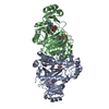
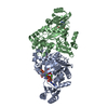
| Entry | Database: PDB / ID: 3eth | ||||||
|---|---|---|---|---|---|---|---|
| Title | Crystal structure of E. coli Purk in complex with MgATP | ||||||
 Components Components | Phosphoribosylaminoimidazole carboxylase ATPase subunit | ||||||
 Keywords Keywords | LYASE / ATP-grasp / purine biosynthesis / antimicrobial / ATP-binding / Decarboxylase / Nucleotide-binding | ||||||
| Function / homology |  Function and homology information Function and homology information5-(carboxyamino)imidazole ribonucleotide synthase / 5-(carboxyamino)imidazole ribonucleotide synthase activity / phosphoribosylaminoimidazole carboxylase activity / 'de novo' IMP biosynthetic process / ATP binding / metal ion binding / cytosol Similarity search - Function | ||||||
| Biological species |  | ||||||
| Method |  X-RAY DIFFRACTION / SYNCHROTRON / MOLECULAR REPLACEMENT / molecular replacement / Resolution: 1.6 Å X-RAY DIFFRACTION / SYNCHROTRON / MOLECULAR REPLACEMENT / molecular replacement / Resolution: 1.6 Å | ||||||
 Authors Authors | Holden, H.M. / Thoden, J.B. | ||||||
 Citation Citation | Journal: Biochemistry / Year: 2008 Title: Structural analysis of the active site geometry of N(5)-Carboxyaminoimidazole ribonucleotide synthetase from Escherichia coli. Authors: Thoden, J.B. / Holden, H.M. / Firestine, S.M. | ||||||
| History |
|
- Structure visualization
| Structure viewer | Molecule: MolmilJmol/JSmol |
|---|
- Downloads & links
-Download
| PDBx/mmCIF format | 3eth.cif.gz | 169.4 KB | Display | PDBx/mmCIF format |
|---|---|---|---|---|
| PDB format | pdb3eth.ent.gz | 131.4 KB | Display | PDB format |
| PDBx/mmJSON format | 3eth.json.gz | Tree view | PDBx/mmJSON format | |
| Others |  Other downloads Other downloads |
-Validation report
| Arichive directory | https://data.pdbj.org/pub/pdb/validation_reports/et/3ethftp://data.pdbj.org/pub/pdb/validation_reports/et/3eth | HTTPS FTP |
|---|
-Related structure data
| Related structure data | 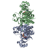 3etjC 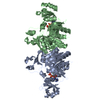 1b6sS C: citing same article ( S: Starting model for refinement |
|---|---|
| Similar structure data |


-Links
 PDBj
PDBj


- Assembly
| Deposited unit | 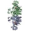
| ||||||||
|---|---|---|---|---|---|---|---|---|---|
| 1 |
| ||||||||
| Unit cell |
|
-Components
| #1: Protein | Mass: 39528.000 Da / Num. of mol.: 2 / Mutation: E61Q, Q205R Source method: isolated from a genetically manipulated source Source: (gene. exp.) References: UniProt: P09029, phosphoribosylaminoimidazole carboxylase #2: Chemical |   Mass: 507.181 Da / Num. of mol.: 2 / Source method: obtained synthetically / Formula: C10H16N5O13P3 / Comment: ATP, energy-carrying molecule*YM Mass: 507.181 Da / Num. of mol.: 2 / Source method: obtained synthetically / Formula: C10H16N5O13P3 / Comment: ATP, energy-carrying molecule*YM#3: Chemical | ChemComp-MG /   Mass: 24.305 Da / Num. of mol.: 4 / Source method: obtained synthetically / Formula: Mg Mass: 24.305 Da / Num. of mol.: 4 / Source method: obtained synthetically / Formula: Mg#4: Water | ChemComp-HOH / |  Mass: 18.015 Da / Num. of mol.: 690 / Source method: isolated from a natural source / Formula: H2O Mass: 18.015 Da / Num. of mol.: 690 / Source method: isolated from a natural source / Formula: H2O |
|---|
-Experimental details
-Experiment
| Experiment | Method: X-RAY DIFFRACTION / Number of used crystals: 1 |
|---|
- Sample preparation
| Crystal | Density Matthews: 2.32 Å3/Da / Density % sol: 46.9 % |
|---|---|
| Crystal grow | Temperature: 298 K / Method: vapor diffusion, hanging drop / pH: 7.5 Details: 20-25% monomethylether poly(ethylene glycol) 5000, 10 mM MgATP, 10 mM aminopyrazole ribonucleotide, pH 7.5, VAPOR DIFFUSION, HANGING DROP, temperature 298K |
-Data collection
| Diffraction | Mean temperature: 100 K |
|---|---|
| Diffraction source | Source: SYNCHROTRON / Site: APS  / Beamline: 19-ID / Wavelength: 0.98 Å / Beamline: 19-ID / Wavelength: 0.98 Å |
| Detector | Type: SBC-3 / Detector: CCD / Date: Dec 6, 2006 |
| Radiation | Protocol: SINGLE WAVELENGTH / Monochromatic (M) / Laue (L): M / Scattering type: x-ray |
| Radiation wavelength | Wavelength: 0.98 Å / Relative weight: 1 |
| Reflection | Resolution: 1.6→30 Å / Num. all: 87837 / Num. obs: 87837 / % possible obs: 93.8 % / Observed criterion σ(F): 0 / Observed criterion σ(I): 0 / Redundancy: 2.3 % / Rmerge(I) obs: 0.033 / Rsym value: 0.033 / Net I/σ(I): 29.5 |
| Reflection shell | Resolution: 1.6→1.66 Å / Redundancy: 1.8 % / Rmerge(I) obs: 0.098 / Mean I/σ(I) obs: 6.4 / Num. unique all: 7649 / Rsym value: 0.098 / % possible all: 81.9 |
-Phasing
| Phasing | Method: molecular replacement |
|---|
- Processing
| Software |
| ||||||||||||||||||||||||||||||||||||
|---|---|---|---|---|---|---|---|---|---|---|---|---|---|---|---|---|---|---|---|---|---|---|---|---|---|---|---|---|---|---|---|---|---|---|---|---|---|
| Refinement | Method to determine structure: MOLECULAR REPLACEMENT Starting model: pdb entry 1b6s Resolution: 1.6→30 Å / Occupancy max: 1 / Occupancy min: 0.5 / Cross valid method: THROUGHOUT / σ(F): 0 / σ(I): 0 / Stereochemistry target values: Engh & Huber
| ||||||||||||||||||||||||||||||||||||
| Displacement parameters | Biso max: 99.85 Å2 / Biso mean: 27.439 Å2 / Biso min: 6.97 Å2 | ||||||||||||||||||||||||||||||||||||
| Refinement step | Cycle: LAST / Resolution: 1.6→30 Å
|
