 ムービー
ムービー コントローラー


+ データを開く

- 基本情報
基本情報
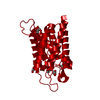
| 登録情報 | データベース: PDB / ID: 1ih5 | |||||||||
|---|---|---|---|---|---|---|---|---|---|---|
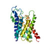
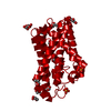
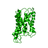
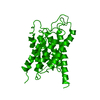
| タイトル | CRYSTAL STRUCTURE OF AQUAPORIN-1 | |||||||||
 要素 要素 | AQUAPORIN-1 | |||||||||
 キーワード キーワード | MEMBRANE PROTEIN / WATER CHANNEL / TWO-DIMENSIONAL CRYSTAL | |||||||||
| 機能・相同性 |  機能・相同性情報 機能・相同性情報metanephric descending thin limb development / metanephric proximal straight tubule development / metanephric proximal convoluted tubule segment 2 development / metanephric glomerulus vasculature development / hydrogen peroxide channel activity / nitric oxide transmembrane transporter activity / lipid digestion / cellular response to salt stress / renal water transport / corticotropin secretion ...metanephric descending thin limb development / metanephric proximal straight tubule development / metanephric proximal convoluted tubule segment 2 development / metanephric glomerulus vasculature development / hydrogen peroxide channel activity / nitric oxide transmembrane transporter activity / lipid digestion / cellular response to salt stress / renal water transport / corticotropin secretion / carbon dioxide transmembrane transport / renal water absorption / carbon dioxide transmembrane transporter activity / glycerol transmembrane transporter activity / secretory granule organization / water transmembrane transporter activity / Passive transport by Aquaporins / cerebrospinal fluid secretion / positive regulation of saliva secretion / pancreatic juice secretion / establishment or maintenance of actin cytoskeleton polarity / lateral ventricle development / glycerol transmembrane transport / cellular response to mercury ion / intracellularly cGMP-activated cation channel activity / potassium ion transmembrane transporter activity / intracellular water homeostasis / water transport / transepithelial water transport / water channel activity / ammonium transmembrane transport / ankyrin-1 complex / ammonium channel activity / glomerular filtration / camera-type eye morphogenesis / fibroblast migration / multicellular organismal-level water homeostasis / cellular homeostasis / cellular hyperosmotic response / cell volume homeostasis / hyperosmotic response / odontogenesis / positive regulation of fibroblast migration / : / nitric oxide transport / brush border / transmembrane transporter activity / cellular response to dexamethasone stimulus / potassium channel activity / renal water homeostasis / ephrin receptor binding / cellular response to retinoic acid / sensory perception of pain / cellular response to nitric oxide / basal plasma membrane / cellular response to copper ion / cellular response to cAMP / carbon dioxide transport / brush border membrane / wound healing / establishment of localization in cell / Erythrocytes take up oxygen and release carbon dioxide / Erythrocytes take up carbon dioxide and release oxygen / cellular response to mechanical stimulus / sarcolemma / potassium ion transport / cellular response to hydrogen peroxide / positive regulation of fibroblast proliferation / positive regulation of angiogenesis / apical part of cell / cellular response to UV / Vasopressin regulates renal water homeostasis via Aquaporins / nuclear membrane / defense response to Gram-negative bacterium / cellular response to hypoxia / basolateral plasma membrane / apical plasma membrane / axon / negative regulation of apoptotic process / extracellular exosome / identical protein binding / nucleus / plasma membrane / cytoplasm 類似検索 - 分子機能 | |||||||||
| 生物種 |  Homo sapiens (ヒト) Homo sapiens (ヒト) | |||||||||
| 手法 | 電子線結晶学 / クライオ電子顕微鏡法 / 解像度: 3.7 Å | |||||||||
 データ登録者 データ登録者 | Ren, G. / Reddy, V.S. / Cheng, A. / Melnyk, P. / Mitra, A.K. | |||||||||
 引用 引用 | ジャーナル: Proc Natl Acad Sci U S A / 年: 2001 タイトル: Visualization of a water-selective pore by electron crystallography in vitreous ice. 著者: G Ren / V S Reddy / A Cheng / P Melnyk / A K Mitra /  要旨: The water-selective pathway through the aquaporin-1 membrane channel has been visualized by fitting an atomic model to a 3.7-A resolution three-dimensional density map. This map was determined by ...The water-selective pathway through the aquaporin-1 membrane channel has been visualized by fitting an atomic model to a 3.7-A resolution three-dimensional density map. This map was determined by analyzing images and electron diffraction patterns of lipid-reconstituted two-dimensional crystals of aquaporin-1 preserved in vitrified buffer in the absence of any additive. The aqueous pathway is characterized by a size-selective pore that is approximately 4.0 +/- 0.5A in diameter, spans a length of approximately 18A, and bends by approximately 25 degrees as it traverses the bilayer. This narrow pore is connected by wide, funnel-shaped openings at the extracellular and cytoplasmic faces. The size-selective pore is outlined mostly by hydrophobic residues, resulting in a relatively inert pathway conducive to diffusion-limited water flow. The apex of the curved pore is close to the locations of the in-plane pseudo-2-fold symmetry axis that relates the N- and C-terminal halves and the conserved, functionally important N76 and N192 residues. #1: ジャーナル: J.Mol.Biol. / 年: 2000タイトル: Three-Dimensional Fold of the Human Aqp1 Water Channel Determined at 4 A Resolution by Electron Crystallography of Two-Dimensional Crystals Embedded in Ice 著者: Ren, G. / Cheng, A. / Reddy, V. / Melnyk, P. / Mitra, A.K. #2: ジャーナル: J.Struct.Biol. / 年: 2000タイトル: Polymorphism in the Packing of Aquaporin-1 Tetramers in 2-D Crystals 著者: Ren, G. / Cheng, A. / Melnyk, P. / Mitra, A.K. #3: ジャーナル: Nature / 年: 1997タイトル: Three-Dimensional Organization of a Human Water Channel 著者: Cheng, A. / Van Hoek, A.N. / Yeager, M. / Verkman, A.S. / Mitra, A.K. | |||||||||
| 履歴 |
|
- 構造の表示
構造の表示
| ムービー |
ムービービューア |
|---|---|
| 構造ビューア | 分子: MolmilJmol/JSmol |
- ダウンロードとリンク
-ダウンロード
| PDBx/mmCIF形式 | 1ih5.cif.gz | 48.3 KB | 表示 | PDBx/mmCIF形式 |
|---|---|---|---|---|
| PDB形式 | pdb1ih5.ent.gz | 34.5 KB | 表示 | PDB形式 |
| PDBx/mmJSON形式 | 1ih5.json.gz | ツリー表示 | PDBx/mmJSON形式 | |
| その他 |  その他のダウンロード その他のダウンロード |
-検証レポート
| 文書・要旨 | 1ih5_validation.pdf.gz | 359.6 KB | 表示 | wwPDB検証レポート |
|---|---|---|---|---|
| 文書・詳細版 | 1ih5_full_validation.pdf.gz | 381.5 KB | 表示 | |
| XML形式データ | 1ih5_validation.xml.gz | 8.3 KB | 表示 | |
| CIF形式データ | 1ih5_validation.cif.gz | 11.6 KB | 表示 | |
| アーカイブディレクトリ | https://data.pdbj.org/pub/pdb/validation_reports/ih/1ih5ftp://data.pdbj.org/pub/pdb/validation_reports/ih/1ih5 | HTTPS FTP |
-関連構造データ
-リンク
 PDBj
PDBj

- 集合体
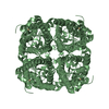
集合体
| 登録構造単位 | 
| ||||||||
|---|---|---|---|---|---|---|---|---|---|
| 1 | 
| ||||||||
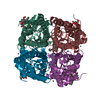
| 単位格子 |
|
-要素
| #1: タンパク質 | 分子量: 28549.914 Da / 分子数: 1 / 由来タイプ: 天然 / 由来: (天然) Homo sapiens (ヒト) / Cell: RED-BLOOD CELL / 細胞内の位置: MEMBRANE / 参照: UniProt: P29972 |
|---|
-実験情報
-実験
| 実験 | 手法: 電子線結晶学 |
|---|---|
| EM実験 | 試料の集合状態: 2D ARRAY / 3次元再構成法: 電子線結晶学 |
- 試料調製
試料調製
| 構成要素 | 名称: aquaporin / タイプ: COMPLEX | ||||||||||||||||||||||||||||||||||||||||||||||||
|---|---|---|---|---|---|---|---|---|---|---|---|---|---|---|---|---|---|---|---|---|---|---|---|---|---|---|---|---|---|---|---|---|---|---|---|---|---|---|---|---|---|---|---|---|---|---|---|---|---|
| 試料 | 包埋: NO / シャドウイング: NO / 染色: NO / 凍結: YES | ||||||||||||||||||||||||||||||||||||||||||||||||
| 結晶化 | *PLUS pH: 7.2 / 手法: unknown | ||||||||||||||||||||||||||||||||||||||||||||||||
| 溶液の組成 | *PLUS
|
-データ収集
| 顕微鏡 | モデル: FEI/PHILIPS CM200FEG |
|---|---|
| 電子銃 | 電子線源:  FIELD EMISSION GUN / 照射モード: FLOOD BEAM FIELD EMISSION GUN / 照射モード: FLOOD BEAM |
| 電子レンズ | モード: DIFFRACTION |
| 検出器 | 日付: 1999年1月1日 |
| 放射 | プロトコル: SINGLE WAVELENGTH / 単色(M)・ラウエ(L): M / 散乱光タイプ: electron |
| 放射波長 | 相対比: 1 |
| 反射 | Biso Wilson estimate: 38.1 Å2 |
| 反射 | *PLUS 最高解像度: 3.7 Å / Num. obs: 19839 / % possible obs: 63 % / Num. measured all: 48037 |
- 解析
解析
| ソフトウェア | 名称: X-PLOR / 分類: 精密化 | ||||||||||||||||||||
|---|---|---|---|---|---|---|---|---|---|---|---|---|---|---|---|---|---|---|---|---|---|
| 3次元再構成 | 解像度: 3.7 Å / 解像度の算出法: DIFFRACTION PATTERN/LAYERLINES | ||||||||||||||||||||
| 精密化 | 解像度: 3.7→24 Å / Isotropic thermal model: ISOTROPIC / 交差検証法: THROUGHOUT / σ(F): 2 詳細: POSITIONAL REFINEMENT USING X-PLOR. REFINEMENT MONITORED BY RFREE AND PHIFREE (<| CALCLD. PHASE - EXPTL. OBSERVED PHASE|>). PSEUDO 2-FOLD SYMMETRY IN THE PLANE OF THE BILAYER RELATES THE N- AND C-TERMINAL HALVES
| ||||||||||||||||||||
| 原子変位パラメータ | Biso mean: 47.8 Å2 | ||||||||||||||||||||
| 精密化ステップ | サイクル: LAST / 解像度: 3.7→24 Å
|
