 Movie
Movie Controller


+ Open data

- Basic information
| Entry | Database: PDB / ID: 1ie4 | |||||||||
|---|---|---|---|---|---|---|---|---|---|---|
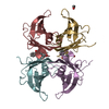
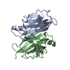
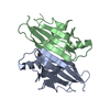
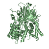
| Title | RAT TRANSTHYRETIN COMPLEX WITH THYROXINE (T4) | |||||||||
 Components Components | TRANSTHYRETIN | |||||||||
 Keywords Keywords | TRANSPORT PROTEIN / TRANSPORT OF THYROID HORMONES / RAT TRANSTHYRETIN / PREALBUMIN / THYROXINE COMPLEX | |||||||||
| Function / homology |  Function and homology information Function and homology informationThe canonical retinoid cycle in rods (twilight vision) / Retinoid metabolism and transport / thyroid hormone metabolic process / thyroid hormone binding / Neutrophil degranulation / negative regulation of glomerular filtration / small molecule binding / purine nucleobase metabolic process / hormone binding / phototransduction, visible light ...The canonical retinoid cycle in rods (twilight vision) / Retinoid metabolism and transport / thyroid hormone metabolic process / thyroid hormone binding / Neutrophil degranulation / negative regulation of glomerular filtration / small molecule binding / purine nucleobase metabolic process / hormone binding / phototransduction, visible light / molecular sequestering activity / retinoid metabolic process / hormone activity / protein-containing complex binding / protein-containing complex / : / extracellular region / identical protein binding Similarity search - Function | |||||||||
| Biological species |  | |||||||||
| Method |  X-RAY DIFFRACTION / FOURIER SYNTHESIS / Resolution: 2.5 Å X-RAY DIFFRACTION / FOURIER SYNTHESIS / Resolution: 2.5 Å | |||||||||
 Authors Authors | Wojtczak, A. | |||||||||
 Citation Citation | Journal: Acta Crystallogr.,Sect.D / Year: 2001 Title: Structure of rat transthyretin (rTTR) complex with thyroxine at 2.5 A resolution: first non-biased insight into thyroxine binding reveals different hormone orientation in two binding sites. Authors: Wojtczak, A. / Cody, V. / Luft, J.R. / Pangborn, W. #1: Journal: Acta Crystallogr.,Sect.D / Year: 1996Title: Structures of Human Transthyretin Complexed with Thyroxine at 2.0 ? Resolution and 3',5'-Dinitro-N-acetyl-L-thyronine at 2.2 ? Resolution Authors: Wojtczak, A. / Cody, V. / Luft, J. / Pangborn, W. #2: Journal: ACTA BIOCHIM.POL. / Year: 1997Title: Crystal Structure of Rat Transthyretin at 2.5 A Resolution: First Report on a Unique Tetrameric Structure. Authors: Wojtczak, A. | |||||||||
| History |
|
- Structure visualization
| Structure viewer | Molecule: MolmilJmol/JSmol |
|---|
- Downloads & links
-Download
| PDBx/mmCIF format | 1ie4.cif.gz | 106.6 KB | Display | PDBx/mmCIF format |
|---|---|---|---|---|
| PDB format | pdb1ie4.ent.gz | 82.6 KB | Display | PDB format |
| PDBx/mmJSON format | 1ie4.json.gz | Tree view | PDBx/mmJSON format | |
| Others |  Other downloads Other downloads |
-Validation report
| Arichive directory | https://data.pdbj.org/pub/pdb/validation_reports/ie/1ie4ftp://data.pdbj.org/pub/pdb/validation_reports/ie/1ie4 | HTTPS FTP |
|---|
-Related structure data
| Related structure data |  1gkeS S: Starting model for refinement |
|---|---|
| Similar structure data |


-Links
 PDBj
PDBj


- Assembly
| Deposited unit | 
| ||||||||
|---|---|---|---|---|---|---|---|---|---|
| 1 |
| ||||||||
| Unit cell |
|
-Components
| #1: Protein | Mass: 13614.147 Da / Num. of mol.: 4 / Source method: isolated from a natural source Details: THIS SEQUENCE OCCURS NATURALLY IN RAT; PURIFIED FROM NATURAL SOURCE (SERUM) Source: (natural) #2: Chemical |   Mass: 776.870 Da / Num. of mol.: 2 / Source method: obtained synthetically / Formula: C15H11I4NO4 / Comment: hormone*YM Mass: 776.870 Da / Num. of mol.: 2 / Source method: obtained synthetically / Formula: C15H11I4NO4 / Comment: hormone*YM#3: Water | ChemComp-HOH / |  Mass: 18.015 Da / Num. of mol.: 172 / Source method: isolated from a natural source / Formula: H2O Mass: 18.015 Da / Num. of mol.: 172 / Source method: isolated from a natural source / Formula: H2O |
|---|
-Experimental details
-Experiment
| Experiment | Method: X-RAY DIFFRACTION / Number of used crystals: 1 |
|---|
- Sample preparation
| Crystal | Density Matthews: 2.472 Å3/Da / Density % sol: 50.5 % | ||||||||||||||||||||||||||||||
|---|---|---|---|---|---|---|---|---|---|---|---|---|---|---|---|---|---|---|---|---|---|---|---|---|---|---|---|---|---|---|---|
| Crystal grow | Temperature: 293 K / Method: vapor diffusion, hanging drop / pH: 5 Details: Ammonium sulfate, acetate buffer, pH 5.0, VAPOR DIFFUSION, HANGING DROP, temperature 293K | ||||||||||||||||||||||||||||||
| Crystal grow | *PLUS | ||||||||||||||||||||||||||||||
| Components of the solutions | *PLUS
|
-Data collection
| Diffraction | Mean temperature: 293 K |
|---|---|
| Diffraction source | Source: ROTATING ANODE / Type: RIGAKU RU200 / Wavelength: 1.5418 Å |
| Detector | Type: RIGAKU RAXIS II / Detector: IMAGE PLATE / Date: Oct 1, 1994 / Details: Collimator |
| Radiation | Monochromator: GRAPHITE / Protocol: SINGLE WAVELENGTH / Monochromatic (M) / Laue (L): M / Scattering type: x-ray |
| Radiation wavelength | Wavelength: 1.5418 Å / Relative weight: 1 |
| Reflection | Resolution: 2.5→82.5 Å / Num. all: 18890 / Num. obs: 18890 / % possible obs: 93.8 % / Observed criterion σ(F): 0 / Observed criterion σ(I): 0 / Redundancy: 4.4 % / Biso Wilson estimate: 17.4 Å2 / Rmerge(I) obs: 0.106 / Net I/σ(I): 6 |
| Reflection shell | Resolution: 2.5→2.6 Å / Redundancy: 2.14 % / Rmerge(I) obs: 0.35 / Mean I/σ(I) obs: 2.19 / Num. unique all: 1786 / % possible all: 82.2 |
| Reflection | *PLUS Highest resolution: 2.5 Å / Lowest resolution: 82.5 Å / Num. obs: 16558 / % possible obs: 83.5 % / Num. measured all: 83761 |
| Reflection shell | *PLUS Mean I/σ(I) obs: 2.1 |
- Processing
| Software |
| ||||||||||||||||||||||||||||||||||||
|---|---|---|---|---|---|---|---|---|---|---|---|---|---|---|---|---|---|---|---|---|---|---|---|---|---|---|---|---|---|---|---|---|---|---|---|---|---|
| Refinement | Method to determine structure: FOURIER SYNTHESIS Starting model: PDB ENTRY 1GKE Resolution: 2.5→12 Å / Rfactor Rfree error: 0.008 / Data cutoff high absF: 177534.13 / Data cutoff low absF: 0 / Isotropic thermal model: RESTRAINED / Cross valid method: THROUGHOUT / σ(F): 2.5 / Stereochemistry target values: Engh & Huber
| ||||||||||||||||||||||||||||||||||||
| Solvent computation | Solvent model: FLAT MODEL / Bsol: 17.32 Å2 / ksol: 0.317 e/Å3 | ||||||||||||||||||||||||||||||||||||
| Displacement parameters | Biso mean: 17.8 Å2
| ||||||||||||||||||||||||||||||||||||
| Refine analyze |
| ||||||||||||||||||||||||||||||||||||
| Refinement step | Cycle: LAST / Resolution: 2.5→12 Å
| ||||||||||||||||||||||||||||||||||||
| Refine LS restraints |
| ||||||||||||||||||||||||||||||||||||
| LS refinement shell | Resolution: 2.5→2.66 Å / Rfactor Rfree error: 0.032 / Total num. of bins used: 6
| ||||||||||||||||||||||||||||||||||||
| Xplor file |
| ||||||||||||||||||||||||||||||||||||
| Refine LS restraints | *PLUS
|
