 Movie
Movie Controller

[English] 日本語
 Yorodumi
- PDB-1i8f: THE CRYSTAL STRUCTURE OF A HEPTAMERIC ARCHAEAL SM PROTEIN: IMPLIC... -
+ Open data

- Basic information
| Entry | Database: PDB / ID: 1i8f | ||||||
|---|---|---|---|---|---|---|---|
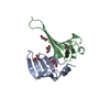
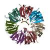
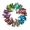
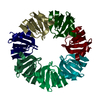
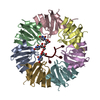
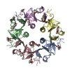
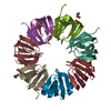
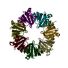
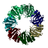
| Title | THE CRYSTAL STRUCTURE OF A HEPTAMERIC ARCHAEAL SM PROTEIN: IMPLICATIONS FOR THE EUKARYOTIC SNRNP CORE | ||||||
 Components Components | PUTATIVE SNRNP SM-LIKE PROTEIN | ||||||
 Keywords Keywords | STRUCTURAL GENOMICS / beta barrel-like SmAP monomers form 35-stranded beta-sheet in the heptamer | ||||||
| Function / homology |  Function and homology information Function and homology information | ||||||
| Biological species |   Pyrobaculum aerophilum (archaea) Pyrobaculum aerophilum (archaea) | ||||||
| Method |  X-RAY DIFFRACTION / SYNCHROTRON / MAD / Resolution: 1.75 Å X-RAY DIFFRACTION / SYNCHROTRON / MAD / Resolution: 1.75 Å | ||||||
 Authors Authors | Mura, C. / Cascio, D. / Sawaya, M.R. / Eisenberg, D. | ||||||
 Citation Citation | Journal: Proc.Natl.Acad.Sci.USA / Year: 2001 Title: The crystal structure of a heptameric archaeal Sm protein: Implications for the eukaryotic snRNP core. Authors: Mura, C. / Cascio, D. / Sawaya, M.R. / Eisenberg, D.S. | ||||||
| History |
|
- Structure visualization
| Structure viewer | Molecule: MolmilJmol/JSmol |
|---|
- Downloads & links
-Download
| PDBx/mmCIF format | 1i8f.cif.gz | 107.8 KB | Display | PDBx/mmCIF format |
|---|---|---|---|---|
| PDB format | pdb1i8f.ent.gz | 85 KB | Display | PDB format |
| PDBx/mmJSON format | 1i8f.json.gz | Tree view | PDBx/mmJSON format | |
| Others |  Other downloads Other downloads |
-Validation report
| Summary document | 1i8f_validation.pdf.gz | 473.2 KB | Display | wwPDB validaton report |
|---|---|---|---|---|
| Full document | 1i8f_full_validation.pdf.gz | 485.3 KB | Display | |
| Data in XML | 1i8f_validation.xml.gz | 22 KB | Display | |
| Data in CIF | 1i8f_validation.cif.gz | 30.7 KB | Display | |
| Arichive directory | https://data.pdbj.org/pub/pdb/validation_reports/i8/1i8fftp://data.pdbj.org/pub/pdb/validation_reports/i8/1i8f | HTTPS FTP |
-Related structure data
| Related structure data | |
|---|---|
| Similar structure data |


-Links
 PDBj
PDBj

- Assembly
| Deposited unit | 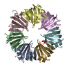
| ||||||||
|---|---|---|---|---|---|---|---|---|---|
| 1 |
| ||||||||
| Unit cell |
| ||||||||
| Details | The contents of one asymmetric unit (i.e. a heptamer) most likely correspond to the biologically relevant species for this organism. |
-Components
| #1: Protein | Mass: 8937.272 Da / Num. of mol.: 7 Source method: isolated from a genetically manipulated source Source: (gene. exp.) Pyrobaculum aerophilum (archaea) / Species (production host): Escherichia coli / Production host:  #2: Chemical | ChemComp-GOL /   Mass: 92.094 Da / Num. of mol.: 5 / Source method: obtained synthetically / Formula: C3H8O3 Mass: 92.094 Da / Num. of mol.: 5 / Source method: obtained synthetically / Formula: C3H8O3#3: Water | ChemComp-HOH / |  Mass: 18.015 Da / Num. of mol.: 130 / Source method: isolated from a natural source / Formula: H2O Mass: 18.015 Da / Num. of mol.: 130 / Source method: isolated from a natural source / Formula: H2O |
|---|
-Experimental details
-Experiment
| Experiment | Method: X-RAY DIFFRACTION / Number of used crystals: 1 |
|---|
- Sample preparation
| Crystal | Density Matthews: 2.38 Å3/Da / Density % sol: 48.34 % |
|---|---|
| Crystal grow | Temperature: 293 K / Method: vapor diffusion, hanging drop / pH: 8.3 Details: PEG-4000, acetate, glycerol, pH 8.3, VAPOR DIFFUSION, HANGING DROP, temperature 293.0K |
| Crystal grow | *PLUS Method: unknown |
-Data collection
| Diffraction | Mean temperature: 105 K |
|---|---|
| Diffraction source | Source: SYNCHROTRON / Site: NSLS  / Beamline: X8C / Wavelength: 0.9794 Å / Beamline: X8C / Wavelength: 0.9794 Å |
| Detector | Type: ADSC QUANTUM 4 / Detector: CCD / Date: Nov 5, 2000 Details: collimating mirror optics, double-slit monochromator |
| Radiation | Monochromator: Si 111 CHANNEL / Protocol: SINGLE WAVELENGTH / Monochromatic (M) / Laue (L): M / Scattering type: x-ray |
| Radiation wavelength | Wavelength: 0.9794 Å / Relative weight: 1 |
| Reflection | Resolution: 1.71→100 Å / Num. all: 62547 / Num. obs: 62547 / % possible obs: 98.5 % / Observed criterion σ(F): 0 / Observed criterion σ(I): -3 / Redundancy: 6.48 % / Rmerge(I) obs: 0.04 / Net I/σ(I): 44.4 |
| Reflection shell | Resolution: 1.71→1.77 Å / Rmerge(I) obs: 0.723 / % possible all: 97.2 |
| Reflection | *PLUS Rmerge(I) obs: 0.04 |
- Processing
| Software |
| |||||||||||||||||||||||||
|---|---|---|---|---|---|---|---|---|---|---|---|---|---|---|---|---|---|---|---|---|---|---|---|---|---|---|
| Refinement | Method to determine structure: MAD / Resolution: 1.75→20 Å / Isotropic thermal model: anisotropic / σ(F): 0 / σ(I): 0 / Stereochemistry target values: Engh & Huber Details: Each of the seven Sm monomers per a.u. were refined independently in CNS since imposition of restraints or constraints hindered the refinement.
| |||||||||||||||||||||||||
| Displacement parameters | Biso mean: 38.5 Å2 | |||||||||||||||||||||||||
| Refine analyze |
| |||||||||||||||||||||||||
| Refinement step | Cycle: LAST / Resolution: 1.75→20 Å
| |||||||||||||||||||||||||
| Refine LS restraints |
| |||||||||||||||||||||||||
| Software | *PLUS Name: CNS / Version: 1 / Classification: refinement | |||||||||||||||||||||||||
| Refinement | *PLUS Lowest resolution: 20 Å / σ(F): 0 / % reflection Rfree: 5 % / Rfactor obs: 0.235 | |||||||||||||||||||||||||
| Solvent computation | *PLUS | |||||||||||||||||||||||||
| Displacement parameters | *PLUS Biso mean: 38.5 Å2 | |||||||||||||||||||||||||
| Refine LS restraints | *PLUS
|
