 Movie
Movie Controller

[English] 日本語
 Yorodumi
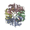
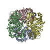
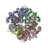
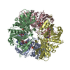
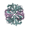
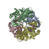
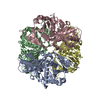
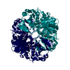
- PDB-1i32: LEISHMANIA MEXICANA GLYCERALDEHYDE-3-PHOSPHATE DEHYDROGENASE IN C... -
+ Open data

- Basic information
| Entry | Database: PDB / ID: 1i32 | ||||||
|---|---|---|---|---|---|---|---|
| Title | LEISHMANIA MEXICANA GLYCERALDEHYDE-3-PHOSPHATE DEHYDROGENASE IN COMPLEX WITH INHIBITORS | ||||||
 Components Components | GLYCERALDEHYDE 3-PHOSPHATE DEHYDROGENASE | ||||||
 Keywords Keywords | OXIDOREDUCTASE / enzyme / dehydrogenase | ||||||
| Function / homology |  Function and homology information Function and homology informationglyceraldehyde-3-phosphate dehydrogenase (phosphorylating) / glyceraldehyde-3-phosphate dehydrogenase (NAD+) (phosphorylating) activity / glycosome / glycolytic process / glucose metabolic process / NAD binding / NADP binding / cytosol Similarity search - Function | ||||||
| Biological species |   Leishmania mexicana (eukaryote) Leishmania mexicana (eukaryote) | ||||||
| Method |  X-RAY DIFFRACTION / SYNCHROTRON / MOLECULAR REPLACEMENT / Resolution: 2.6 Å X-RAY DIFFRACTION / SYNCHROTRON / MOLECULAR REPLACEMENT / Resolution: 2.6 Å | ||||||
 Authors Authors | Suresh, S. / Bressi, J.C. / Kennedy, K.J. / Verlinde, C.L.M.J. / Gelb, M.H. / Hol, W.G.J. | ||||||
 Citation Citation | Journal: J.Mol.Biol. / Year: 2001 Title: Conformational changes in Leishmania mexicana glyceraldehyde-3-phosphate dehydrogenase induced by designed inhibitors. Authors: Suresh, S. / Bressi, J.C. / Kennedy, K.J. / Verlinde, C.L. / Gelb, M.H. / Hol, W.G. | ||||||
| History |
|
- Structure visualization
| Structure viewer | Molecule: MolmilJmol/JSmol |
|---|
- Downloads & links
-Download
| PDBx/mmCIF format | 1i32.cif.gz | 424.1 KB | Display | PDBx/mmCIF format |
|---|---|---|---|---|
| PDB format | pdb1i32.ent.gz | 348.4 KB | Display | PDB format |
| PDBx/mmJSON format | 1i32.json.gz | Tree view | PDBx/mmJSON format | |
| Others |  Other downloads Other downloads |
-Validation report
| Summary document | 1i32_validation.pdf.gz | 841.3 KB | Display | wwPDB validaton report |
|---|---|---|---|---|
| Full document | 1i32_full_validation.pdf.gz | 891.3 KB | Display | |
| Data in XML | 1i32_validation.xml.gz | 48.7 KB | Display | |
| Data in CIF | 1i32_validation.cif.gz | 71.1 KB | Display | |
| Arichive directory | https://data.pdbj.org/pub/pdb/validation_reports/i3/1i32ftp://data.pdbj.org/pub/pdb/validation_reports/i3/1i32 | HTTPS FTP |
-Related structure data


-Links
 PDBj
PDBj
- Assembly
| Deposited unit | 
| ||||||||
|---|---|---|---|---|---|---|---|---|---|
| 1 | 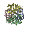
| ||||||||
| 2 | 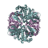
| ||||||||
| Unit cell |
|
-Components
| #1: Protein | Mass: 38953.504 Da / Num. of mol.: 6 Source method: isolated from a genetically manipulated source Source: (gene. exp.) Leishmania mexicana (eukaryote) / Production host:  References: UniProt: Q27890, glyceraldehyde-3-phosphate dehydrogenase (phosphorylating) #2: Chemical | ChemComp-NMD /   Mass: 570.596 Da / Num. of mol.: 6 / Source method: obtained synthetically / Formula: C30H30N6O6 Mass: 570.596 Da / Num. of mol.: 6 / Source method: obtained synthetically / Formula: C30H30N6O6#3: Water | ChemComp-HOH / |  Mass: 18.015 Da / Num. of mol.: 657 / Source method: isolated from a natural source / Formula: H2O Mass: 18.015 Da / Num. of mol.: 657 / Source method: isolated from a natural source / Formula: H2O |
|---|
-Experimental details
-Experiment
| Experiment | Method: X-RAY DIFFRACTION / Number of used crystals: 1 |
|---|
- Sample preparation
| Crystal | Density Matthews: 2.4 Å3/Da / Density % sol: 48.83 % | ||||||||||||||||||||||||||||||||||||||||||||||||||||||
|---|---|---|---|---|---|---|---|---|---|---|---|---|---|---|---|---|---|---|---|---|---|---|---|---|---|---|---|---|---|---|---|---|---|---|---|---|---|---|---|---|---|---|---|---|---|---|---|---|---|---|---|---|---|---|---|
| Crystal grow | Temperature: 298 K / Method: vapor diffusion, sitting drop / pH: 7.25 Details: PEG1000, 100mM TEA, 250mM NaCl, 5mM DTT, 1mM PMSF 15mM HEPES, pH 7.25, VAPOR DIFFUSION, SITTING DROP, temperature 298K | ||||||||||||||||||||||||||||||||||||||||||||||||||||||
| Crystal grow | *PLUS | ||||||||||||||||||||||||||||||||||||||||||||||||||||||
| Components of the solutions | *PLUS
|
-Data collection
| Diffraction | Mean temperature: 100 K |
|---|---|
| Diffraction source | Source: SYNCHROTRON / Site: ALS  / Beamline: 5.0.2 / Wavelength: 1 Å / Beamline: 5.0.2 / Wavelength: 1 Å |
| Detector | Type: ADSC QUANTUM 4 / Detector: CCD / Date: Apr 16, 2000 |
| Radiation | Monochromator: synchrotron / Protocol: SINGLE WAVELENGTH / Monochromatic (M) / Laue (L): M / Scattering type: x-ray |
| Radiation wavelength | Wavelength: 1 Å / Relative weight: 1 |
| Reflection | Resolution: 2.6→15 Å / Num. all: 65878 / Num. obs: 65878 / % possible obs: 94 % / Observed criterion σ(F): 0 / Observed criterion σ(I): 0 / Redundancy: 6.18 % / Biso Wilson estimate: 47.7 Å2 / Rmerge(I) obs: 0.066 / Rsym value: 0.066 / Net I/σ(I): 29.3 |
| Reflection shell | Resolution: 2.6→2.65 Å / Redundancy: 3.4 % / Rmerge(I) obs: 0.289 / % possible all: 71.7 |
| Reflection | *PLUS Lowest resolution: 15 Å / Num. measured all: 407373 |
| Reflection shell | *PLUS % possible obs: 71.7 % / Mean I/σ(I) obs: 4.7 |
- Processing
| Software |
| ||||||||||||||||||||
|---|---|---|---|---|---|---|---|---|---|---|---|---|---|---|---|---|---|---|---|---|---|
| Refinement | Method to determine structure: MOLECULAR REPLACEMENT / Resolution: 2.6→15 Å / σ(F): 0 / σ(I): 0 / Stereochemistry target values: Engh & Huber
| ||||||||||||||||||||
| Refinement step | Cycle: LAST / Resolution: 2.6→15 Å
| ||||||||||||||||||||
| Refine LS restraints |
| ||||||||||||||||||||
| Software | *PLUS Name: CNS / Classification: refinement | ||||||||||||||||||||
| Refinement | *PLUS Highest resolution: 2.6 Å / Lowest resolution: 15 Å / σ(F): 0 / % reflection Rfree: 5 % | ||||||||||||||||||||
| Solvent computation | *PLUS | ||||||||||||||||||||
| Displacement parameters | *PLUS | ||||||||||||||||||||
| LS refinement shell | *PLUS Rfactor Rfree: 0.368 / Rfactor obs: 0.299 |
