[English] 日本語
Yorodumi
- PDB-5c7l: Structure of human testis-specific glyceraldehyde-3-phosphate deh... -

+
Open data


ID or keywords:

Loading...

-
Basic information

Entry
Database: PDB / ID: 5c7l
TitleStructure of human testis-specific glyceraldehyde-3-phosphate dehydrogenase apo form
ComponentsGlyceraldehyde-3-phosphate dehydrogenase, testis-specificGlyceraldehyde 3-phosphate dehydrogenase
Keywords
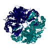
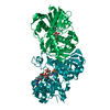
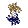
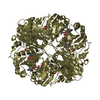
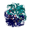
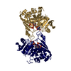
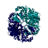
OXIDOREDUCTASE / human sperm GAPDH metabolic enzyme sperm specific
Function / homology
Function and homology information


flagellated sperm motility / glyceraldehyde-3-phosphate dehydrogenase (phosphorylating) / glyceraldehyde-3-phosphate dehydrogenase (NAD+) (phosphorylating) activity / Gluconeogenesis / Glycolysis / Association of TriC/CCT with target proteins during biosynthesis / positive regulation of glycolytic process / glycolytic process / glucose metabolic process / NAD binding ...flagellated sperm motility / glyceraldehyde-3-phosphate dehydrogenase (phosphorylating) / glyceraldehyde-3-phosphate dehydrogenase (NAD+) (phosphorylating) activity / Gluconeogenesis / Glycolysis / Association of TriC/CCT with target proteins during biosynthesis / positive regulation of glycolytic process / glycolytic process / glucose metabolic process / NAD binding / NADP binding / nucleus / cytosol
Similarity search - Function
Glyceraldehyde-3-phosphate dehydrogenase, type I / Glyceraldehyde 3-phosphate dehydrogenase, active site / Glyceraldehyde 3-phosphate dehydrogenase active site. / Glyceraldehyde 3-phosphate dehydrogenase, NAD binding domain / Glyceraldehyde 3-phosphate dehydrogenase, NAD(P) binding domain / Glyceraldehyde 3-phosphate dehydrogenase, catalytic domain / Glyceraldehyde/Erythrose phosphate dehydrogenase family / Glyceraldehyde 3-phosphate dehydrogenase, C-terminal domain / Glyceraldehyde 3-phosphate dehydrogenase, NAD binding domain / Dihydrodipicolinate Reductase; domain 2 ...Glyceraldehyde-3-phosphate dehydrogenase, type I / Glyceraldehyde 3-phosphate dehydrogenase, active site / Glyceraldehyde 3-phosphate dehydrogenase active site. / Glyceraldehyde 3-phosphate dehydrogenase, NAD binding domain / Glyceraldehyde 3-phosphate dehydrogenase, NAD(P) binding domain / Glyceraldehyde 3-phosphate dehydrogenase, catalytic domain / Glyceraldehyde/Erythrose phosphate dehydrogenase family / Glyceraldehyde 3-phosphate dehydrogenase, C-terminal domain / Glyceraldehyde 3-phosphate dehydrogenase, NAD binding domain / Dihydrodipicolinate Reductase; domain 2 / Dihydrodipicolinate Reductase; domain 2 / NAD(P)-binding Rossmann-like Domain / NAD(P)-binding domain superfamily / Rossmann fold / 2-Layer Sandwich / 3-Layer(aba) Sandwich / Alpha Beta
Similarity search - Domain/homology
Glyceraldehyde-3-phosphate dehydrogenase, testis-specific
Similarity search - Component
Biological speciesHomo sapiens (human)
MethodX-RAY DIFFRACTION / SYNCHROTRON / Resolution: 1.862 Å
AuthorsBetts, L. / Machius, M. / Danshina, P. / O'Brien, D.
Funding support United States, 1items
OrganizationGrant numberCountry
National Institutes of Health/Eunice Kennedy Shriver National Institute of Child Health & Human Development (NIH/NICHD) United States
CitationJournal: Mol. Hum. Reprod. / Year: 2016
Title: Structural analyses to identify selective inhibitors of glyceraldehyde 3-phosphate dehydrogenase-S, a sperm-specific glycolytic enzyme.
Authors: Danshina, P.V. / Qu, W. / Temple, B.R. / Rojas, R.J. / Miley, M.J. / Machius, M. / Betts, L. / O'Brien, D.A.
History
DepositionJun 24, 2015Deposition site: RCSB / Processing site: RCSB
Revision 1.0Mar 9, 2016Provider: repository / Type: Initial release
Revision 1.1Jun 8, 2016Group: Database references
Revision 1.2Sep 20, 2017Group: Author supporting evidence / Derived calculations / Category: pdbx_audit_support / pdbx_struct_oper_list
Item: _pdbx_audit_support.funding_organization / _pdbx_struct_oper_list.symmetry_operation
Revision 1.3Dec 11, 2019Group: Author supporting evidence / Category: pdbx_audit_support / Item: _pdbx_audit_support.funding_organization

-
Structure visualization

Structure viewerMolecule:
MolmilJmol/JSmol

Downloads & links

-
Assembly

Deposited unit
O: Glyceraldehyde-3-phosphate dehydrogenase, testis-specific
R: Glyceraldehyde-3-phosphate dehydrogenase, testis-specific


Theoretical massNumber of molelcules
Total (without water)73,4782
Polymers73,4782
Non-polymers00
Water6,918384
1
O: Glyceraldehyde-3-phosphate dehydrogenase, testis-specific
R: Glyceraldehyde-3-phosphate dehydrogenase, testis-specific

O: Glyceraldehyde-3-phosphate dehydrogenase, testis-specific
R: Glyceraldehyde-3-phosphate dehydrogenase, testis-specific


Theoretical massNumber of molelcules
Total (without water)146,9554
Polymers146,9554
Non-polymers00
Water724
TypeNameSymmetry operationNumber
identity operation1_555x,y,z1
crystal symmetry operation5_675x-y+1,-y+2,-z+2/31
Buried area15550 Å2
ΔGint-83 kcal/mol
Surface area44700 Å2
MethodPISA
Unit cell
Length a, b, c (Å)86.888, 86.888, 159.606
Angle α, β, γ (deg.)90.000, 90.000, 120.000
Int Tables number152
Space group name H-MP3121
Components on special symmetry positions
IDModelComponents
11O-601-

HOH

21R-539-

HOH

-
Components

#1: Protein Glyceraldehyde-3-phosphate dehydrogenase, testis-specific / Glyceraldehyde 3-phosphate dehydrogenase / Spermatogenic cell-specific glyceraldehyde 3-phosphate dehydrogenase 2 / GAPDH-2 / Spermatogenic ...Spermatogenic cell-specific glyceraldehyde 3-phosphate dehydrogenase 2 / GAPDH-2 / Spermatogenic glyceraldehyde-3-phosphate dehydrogenase


Mass: 36738.758 Da / Num. of mol.: 2 / Fragment: unp residues 74-407
Source method: isolated from a genetically manipulated source
Source: (gene. exp.) Homo sapiens (human) / Gene: GAPDHS, GAPD2, GAPDH2, GAPDS, HSD-35, HSD35 / Production host: Escherichia coli (E. coli)
References: UniProt: O14556, glyceraldehyde-3-phosphate dehydrogenase (phosphorylating)
#2: Water ChemComp-HOH / water / Water


Mass: 18.015 Da / Num. of mol.: 384 / Source method: isolated from a natural source / Formula: H2O

-
Experimental details

-
Experiment

ExperimentMethod: X-RAY DIFFRACTION / Number of used crystals: 1

-
Sample preparation

CrystalDensity Matthews: 2.37 Å3/Da / Density % sol: 48.06 %
Crystal growTemperature: 273 K / Method: vapor diffusion / pH: 6.5
Details: mix 1:1 volume of protein at 10 mg/mL with 0.2 M sodium sulfate, 0.1 M Bis-Tris propane pH 6.5, 20% PEG 3350

-
Data collection

DiffractionMean temperature: 100 K
Diffraction sourceSource: SYNCHROTRON / Site: APS / Beamline: 22-ID / Wavelength: 1.0746 Å
DetectorType: MARMOSAIC 300 mm CCD / Detector: CCD / Date: Apr 10, 2014
RadiationProtocol: SINGLE WAVELENGTH / Monochromatic (M) / Laue (L): M / Scattering type: x-ray
Radiation wavelengthWavelength: 1.0746 Å / Relative weight: 1
ReflectionResolution: 1.86→38.2 Å / Num. obs: 59104 / % possible obs: 99.8 % / Redundancy: 8.7 % / Net I/σ(I): 17.5

-
Processing

Software
NameVersionClassification
SCALEPACKdata scaling
PHENIXrefinement
PDB_EXTRACT3.15data extraction
HKL-2000data reduction
PHENIXphasing
RefinementResolution: 1.862→38.156 Å / SU ML: 0.21 / Cross valid method: FREE R-VALUE / σ(F): 1.35 / Phase error: 23.48 / Stereochemistry target values: ML
RfactorNum. reflection% reflection
Rfree0.227 1996 3.67 %
Rwork0.1865 52425 -
obs0.188 54421 92.01 %
Solvent computationShrinkage radii: 0.9 Å / VDW probe radii: 1.11 Å / Solvent model: FLAT BULK SOLVENT MODEL
Displacement parametersBiso max: 130.51 Å2 / Biso mean: 32.7496 Å2 / Biso min: 8.76 Å2
Refinement stepCycle: final / Resolution: 1.862→38.156 Å
ProteinNucleic acidLigandSolventTotal
Num. atoms5142 0 0 384 5526
Biso mean---33.91 -
Num. residues----672
Refine LS restraints
Refine-IDTypeDev idealNumber
X-RAY DIFFRACTIONf_bond_d0.0135340
X-RAY DIFFRACTIONf_angle_d1.3997274
X-RAY DIFFRACTIONf_chiral_restr0.059814
X-RAY DIFFRACTIONf_plane_restr0.008946
X-RAY DIFFRACTIONf_dihedral_angle_d13.6941962
LS refinement shell

Refine-ID: X-RAY DIFFRACTION / Total num. of bins used: 14

Resolution (Å)Rfactor RfreeNum. reflection RfreeRfactor RworkNum. reflection RworkNum. reflection all% reflection obs (%)
1.8619-1.90840.2597840.22062250233456
1.9084-1.960.28961090.22842736284568
1.96-2.01770.27271250.2373161328679
2.0177-2.08280.30161400.23143562370290
2.0828-2.15730.30451490.23493897404697
2.1573-2.24360.28521540.217440434197100
2.2436-2.34570.25411500.207940374187100
2.3457-2.46940.26981500.200640364186100
2.4694-2.6240.22061550.192640404195100
2.624-2.82660.21951570.191340654222100
2.8266-3.11090.22231550.18640874242100
3.1109-3.56080.22711530.17840964249100
3.5608-4.48510.17871540.156841284282100
4.4851-38.16430.19761610.16324287444899

+
About Yorodumi

-
News

-
Feb 9, 2022. New format data for meta-information of EMDB entries

New format data for meta-information of EMDB entries

  • Version 3 of the EMDB header file is now the official format.
  • The previous official version 1.9 will be removed from the archive.

Related info.:EMDB header

External links:wwPDB to switch to version 3 of the EMDB data model

-
Aug 12, 2020. Covid-19 info

Covid-19 info

URL: https://pdbj.org/emnavi/covid19.php

New page: Covid-19 featured information page in EM Navigator.

Related info.:Covid-19 info / Mar 5, 2020. Novel coronavirus structure data

+
Mar 5, 2020. Novel coronavirus structure data

Novel coronavirus structure data

Related info.:Yorodumi Speices / Aug 12, 2020. Covid-19 info

External links:COVID-19 featured content - PDBj / Molecule of the Month (242):Coronavirus Proteases

+
Jan 31, 2019. EMDB accession codes are about to change! (news from PDBe EMDB page)

EMDB accession codes are about to change! (news from PDBe EMDB page)

  • The allocation of 4 digits for EMDB accession codes will soon come to an end. Whilst these codes will remain in use, new EMDB accession codes will include an additional digit and will expand incrementally as the available range of codes is exhausted. The current 4-digit format prefixed with “EMD-” (i.e. EMD-XXXX) will advance to a 5-digit format (i.e. EMD-XXXXX), and so on. It is currently estimated that the 4-digit codes will be depleted around Spring 2019, at which point the 5-digit format will come into force.
  • The EM Navigator/Yorodumi systems omit the EMD- prefix.

Related info.:Q: What is EMD? / ID/Accession-code notation in Yorodumi/EM Navigator

External links:EMDB Accession Codes are Changing Soon! / Contact to PDBj

+
Jul 12, 2017. Major update of PDB

Major update of PDB

  • wwPDB released updated PDB data conforming to the new PDBx/mmCIF dictionary.
  • This is a major update changing the version number from 4 to 5, and with Remediation, in which all the entries are updated.
  • In this update, many items about electron microscopy experimental information are reorganized (e.g. em_software).
  • Now, EM Navigator and Yorodumi are based on the updated data.

External links:wwPDB Remediation / Enriched Model Files Conforming to OneDep Data Standards Now Available in the PDB FTP Archive

-
Yorodumi

Thousand views of thousand structures

  • Yorodumi is a browser for structure data from EMDB, PDB, SASBDB, etc.
  • This page is also the successor to EM Navigator detail page, and also detail information page/front-end page for Omokage search.
  • The word "yorodu" (or yorozu) is an old Japanese word meaning "ten thousand". "mi" (miru) is to see.

Related info.:EMDB / PDB / SASBDB / Comparison of 3 databanks / Yorodumi Search / Aug 31, 2016. New EM Navigator & Yorodumi / Yorodumi Papers / Jmol/JSmol / Function and homology information / Changes in new EM Navigator and Yorodumi

Read more