 Movie
Movie Controller

[English] 日本語
 Yorodumi
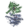
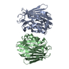
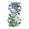
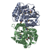
- PDB-1hyn: CRYSTAL STRUCTURE OF THE CYTOPLASMIC DOMAIN OF HUMAN ERYTHROCYTE ... -
+ Open data

- Basic information
| Entry | Database: PDB / ID: 1hyn | ||||||
|---|---|---|---|---|---|---|---|
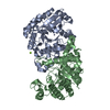
| Title | CRYSTAL STRUCTURE OF THE CYTOPLASMIC DOMAIN OF HUMAN ERYTHROCYTE BAND-3 PROTEIN | ||||||
 Components Components | BAND 3 ANION TRANSPORT PROTEIN | ||||||
 Keywords Keywords | MEMBRANE PROTEIN | ||||||
| Function / homology |  Function and homology information Function and homology informationresponse to increased oxygen levels / pH elevation / Defective SLC4A1 causes hereditary spherocytosis type 4 (HSP4), distal renal tubular acidosis (dRTA) and dRTA with hemolytic anemia (dRTA-HA) / negative regulation of urine volume / Bicarbonate transporters / ankyrin-1 complex / intracellular monoatomic ion homeostasis / monoatomic anion transmembrane transporter activity / plasma membrane phospholipid scrambling / chloride:bicarbonate antiporter activity ...response to increased oxygen levels / pH elevation / Defective SLC4A1 causes hereditary spherocytosis type 4 (HSP4), distal renal tubular acidosis (dRTA) and dRTA with hemolytic anemia (dRTA-HA) / negative regulation of urine volume / Bicarbonate transporters / ankyrin-1 complex / intracellular monoatomic ion homeostasis / monoatomic anion transmembrane transporter activity / plasma membrane phospholipid scrambling / chloride:bicarbonate antiporter activity / solute:inorganic anion antiporter activity / bicarbonate transmembrane transporter activity / bicarbonate transport / monoatomic anion transport / chloride transmembrane transporter activity / chloride transport / ankyrin binding / hemoglobin binding / negative regulation of glycolytic process through fructose-6-phosphate / cortical cytoskeleton / erythrocyte development / protein-membrane adaptor activity / chloride transmembrane transport / regulation of intracellular pH / protein localization to plasma membrane / Erythrocytes take up oxygen and release carbon dioxide / Erythrocytes take up carbon dioxide and release oxygen / transmembrane transport / Z disc / cytoplasmic side of plasma membrane / blood coagulation / blood microparticle / basolateral plasma membrane / protein homodimerization activity / extracellular exosome / membrane / plasma membrane Similarity search - Function | ||||||
| Biological species |  Homo sapiens (human) Homo sapiens (human) | ||||||
| Method |  X-RAY DIFFRACTION / SYNCHROTRON / MAD / Resolution: 2.6 Å X-RAY DIFFRACTION / SYNCHROTRON / MAD / Resolution: 2.6 Å | ||||||
 Authors Authors | Zhang, D. / Kiyatkin, A. / Bolin, J.T. / Low, P.S. | ||||||
 Citation Citation | Journal: Blood / Year: 2000 Title: Crystallographic structure and functional interpretation of the cytoplasmic domain of erythrocyte membrane band 3. Authors: Zhang, D. / Kiyatkin, A. / Bolin, J.T. / Low, P.S. #1: Journal: Proteins / Year: 1995Title: Crystallization and Preliminary X-ray Analysis of the Cytoplasmic Domain of Human Erythrocyte Band 3 Authors: Kiyatkin, A. / Natarajan, P. / Munshi, S. / Minor, W. / Johnson, J.E. / Low, P.S. | ||||||
| History |
|
- Structure visualization
| Structure viewer | Molecule: MolmilJmol/JSmol |
|---|
- Downloads & links
-Download
| PDBx/mmCIF format | 1hyn.cif.gz | 243.7 KB | Display | PDBx/mmCIF format |
|---|---|---|---|---|
| PDB format | pdb1hyn.ent.gz | 198.1 KB | Display | PDB format |
| PDBx/mmJSON format | 1hyn.json.gz | Tree view | PDBx/mmJSON format | |
| Others |  Other downloads Other downloads |
-Validation report
| Arichive directory | https://data.pdbj.org/pub/pdb/validation_reports/hy/1hynftp://data.pdbj.org/pub/pdb/validation_reports/hy/1hyn | HTTPS FTP |
|---|
-Related structure data
| Similar structure data |
|---|


-Links
 PDBj
PDBj- Assembly
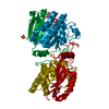
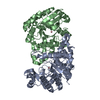
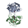
| Deposited unit | 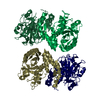
| ||||||||
|---|---|---|---|---|---|---|---|---|---|
| 1 | 
| ||||||||
| 2 | 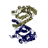
| ||||||||
| 3 | 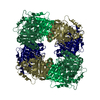
| ||||||||
| Unit cell |
|
-Components
| #1: Protein | Mass: 42576.508 Da / Num. of mol.: 4 Source method: isolated from a genetically manipulated source Source: (gene. exp.) Homo sapiens (human) / Production host:  #2: Water | ChemComp-HOH / |  Mass: 18.015 Da / Num. of mol.: 314 / Source method: isolated from a natural source / Formula: H2O Mass: 18.015 Da / Num. of mol.: 314 / Source method: isolated from a natural source / Formula: H2O |
|---|
-Experimental details
-Experiment
| Experiment | Method: X-RAY DIFFRACTION / Number of used crystals: 1 |
|---|
- Sample preparation
| Crystal | Density Matthews: 2.3 Å3/Da / Density % sol: 46 % | ||||||||||||||||||||||||||||||||||||||||||||||||||||||||||||||||||
|---|---|---|---|---|---|---|---|---|---|---|---|---|---|---|---|---|---|---|---|---|---|---|---|---|---|---|---|---|---|---|---|---|---|---|---|---|---|---|---|---|---|---|---|---|---|---|---|---|---|---|---|---|---|---|---|---|---|---|---|---|---|---|---|---|---|---|---|
| Crystal grow | pH: 4.8 Details: METHOD: SITTING DROP VAPOR DIFFUSION WITH SEEDING. TEMPERATURE: 293 K. RESERVOIR: 50-53% SATURATED AMMONIUM SULFATE, 150mM SODIUM CITRATE PH 4.8. PROTEIN: 7 mg/ml PROTEIN IN 5mM SODIUM ...Details: METHOD: SITTING DROP VAPOR DIFFUSION WITH SEEDING. TEMPERATURE: 293 K. RESERVOIR: 50-53% SATURATED AMMONIUM SULFATE, 150mM SODIUM CITRATE PH 4.8. PROTEIN: 7 mg/ml PROTEIN IN 5mM SODIUM PHOSPHATE PH 6.8, 10 mM SODIUM CHLORIDE. | ||||||||||||||||||||||||||||||||||||||||||||||||||||||||||||||||||
| Crystal grow | *PLUS pH: 7.4 / Method: vapor diffusion, sitting dropDetails: Kiyatkin, A., (1995) Proteins: Struct., Funct., Genet., 22, 293. | ||||||||||||||||||||||||||||||||||||||||||||||||||||||||||||||||||
| Components of the solutions | *PLUS
|
-Data collection
| Diffraction | Mean temperature: 108 K | ||||||||||||
|---|---|---|---|---|---|---|---|---|---|---|---|---|---|
| Diffraction source | Source: SYNCHROTRON / Site: APS  / Beamline: 14-BM-D / Wavelength: 0.9789, 0.9796, 0.9537 / Beamline: 14-BM-D / Wavelength: 0.9789, 0.9796, 0.9537 | ||||||||||||
| Detector | Type: ADSC QUANTUM 1 / Detector: CCD / Date: Sep 23, 1998 / Details: RH-coated toroidal mirror | ||||||||||||
| Radiation | Monochromator: double crystal SI(111) / Protocol: MAD / Monochromatic (M) / Laue (L): M / Scattering type: x-ray | ||||||||||||
| Radiation wavelength |
| ||||||||||||
| Reflection | Resolution: 2.59→50 Å / Num. obs: 46609 / % possible obs: 95.2 % / Observed criterion σ(I): 2 / Redundancy: 3.8 % / Biso Wilson estimate: 46.1 Å2 / Rsym value: 0.052 / Net I/σ(I): 37.16 | ||||||||||||
| Reflection shell | Resolution: 2.59→2.68 Å / Redundancy: 2.8 % / Mean I/σ(I) obs: 18.7 / Rsym value: 0.175 / % possible all: 48.7 | ||||||||||||
| Reflection | *PLUS Lowest resolution: 35 Å / % possible obs: 99.1 % / Rmerge(I) obs: 0.057 | ||||||||||||
| Reflection shell | *PLUS % possible obs: 94.5 % / Rmerge(I) obs: 0.208 |
- Processing
| Software |
| ||||||||||||||||||||||||||||||||||||||||
|---|---|---|---|---|---|---|---|---|---|---|---|---|---|---|---|---|---|---|---|---|---|---|---|---|---|---|---|---|---|---|---|---|---|---|---|---|---|---|---|---|---|
| Refinement | Method to determine structure: MAD / Resolution: 2.6→8 Å / Data cutoff high absF: 0.1 / Data cutoff low absF: 100000 / σ(F): 2
| ||||||||||||||||||||||||||||||||||||||||
| Displacement parameters | Biso mean: 31.3 Å2 | ||||||||||||||||||||||||||||||||||||||||
| Refinement step | Cycle: LAST / Resolution: 2.6→8 Å
| ||||||||||||||||||||||||||||||||||||||||
| Refine LS restraints |
| ||||||||||||||||||||||||||||||||||||||||
| LS refinement shell | Resolution: 2.6→2.71 Å / Total num. of bins used: 8
| ||||||||||||||||||||||||||||||||||||||||
| Xplor file | Serial no: 1 / Param file: parhcsdx.pro | ||||||||||||||||||||||||||||||||||||||||
| Software | *PLUS Name: X-PLOR / Classification: refinement | ||||||||||||||||||||||||||||||||||||||||
| Refinement | *PLUS Highest resolution: 2.6 Å / Lowest resolution: 8 Å / σ(F): 2 / % reflection Rfree: 8.1 % / Rfactor obs: 0.216 / Rfactor Rfree: 0.29 | ||||||||||||||||||||||||||||||||||||||||
| Solvent computation | *PLUS | ||||||||||||||||||||||||||||||||||||||||
| Displacement parameters | *PLUS Biso mean: 31.3 Å2 | ||||||||||||||||||||||||||||||||||||||||
| Refine LS restraints | *PLUS
| ||||||||||||||||||||||||||||||||||||||||
| LS refinement shell | *PLUS Rfactor Rfree: 0.323 / % reflection Rfree: 7.9 % / Rfactor Rwork: 0.29 |
