 Movie
Movie Controller

[English] 日本語
 Yorodumi
- PDB-5gmt: Crystal structure of the marine PL-14 alginate lyase from Aplysia... -
+ Open data

- Basic information
| Entry | Database: PDB / ID: 5gmt | ||||||
|---|---|---|---|---|---|---|---|
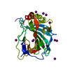
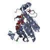
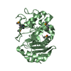
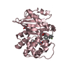
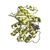
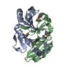
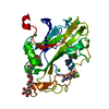
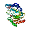
| Title | Crystal structure of the marine PL-14 alginate lyase from Aplysia kurodai | ||||||
 Components Components | Alginate lyase | ||||||
 Keywords Keywords | LYASE / alginate lyase / polysaccharide lyase family 14 / glycosidic bond / beta-D-mannuronic acid / alpha-L-guluronic acid | ||||||
| Function / homology | : / Polysaccharide lyase 14 / lyase activity / Alginate lyase Function and homology information Function and homology information | ||||||
| Biological species |  | ||||||
| Method |  X-RAY DIFFRACTION / SYNCHROTRON / SAD / Resolution: 1.77 Å X-RAY DIFFRACTION / SYNCHROTRON / SAD / Resolution: 1.77 Å | ||||||
 Authors Authors | Qin, H.-M. / Miyakawa, T. / Nakamura, A. / Tanokura, M. | ||||||
| Funding support |  Japan, 1items Japan, 1items
| ||||||
 Citation Citation | Journal: J. Biol. Chem. / Year: 2017 Title: Structure and Polymannuronate Specificity of a Eukaryotic Member of Polysaccharide Lyase Family 14. Authors: Qin, H.M. / Miyakawa, T. / Inoue, A. / Nishiyama, R. / Nakamura, A. / Asano, A. / Sawano, Y. / Ojima, T. / Tanokura, M. | ||||||
| History |
|
- Structure visualization
| Structure viewer | Molecule: MolmilJmol/JSmol |
|---|
- Downloads & links
-Download
| PDBx/mmCIF format | 5gmt.cif.gz | 123.7 KB | Display | PDBx/mmCIF format |
|---|---|---|---|---|
| PDB format | pdb5gmt.ent.gz | 94.9 KB | Display | PDB format |
| PDBx/mmJSON format | 5gmt.json.gz | Tree view | PDBx/mmJSON format | |
| Others |  Other downloads Other downloads |
-Validation report
| Arichive directory | https://data.pdbj.org/pub/pdb/validation_reports/gm/5gmtftp://data.pdbj.org/pub/pdb/validation_reports/gm/5gmt | HTTPS FTP |
|---|
-Related structure data
| Similar structure data |
|---|


-Links
 PDBj
PDBj- Assembly
| Deposited unit | 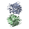
| ||||||||
|---|---|---|---|---|---|---|---|---|---|
| 1 | 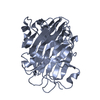
| ||||||||
| 2 | 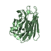
| ||||||||
| Unit cell |
|
-Components
| #1: Protein | Mass: 31169.584 Da / Num. of mol.: 2 / Fragment: UNP residues 29-295 Source method: isolated from a genetically manipulated source Source: (gene. exp.)  References: UniProt: E7FLQ2, mannuronate-specific alginate lyase #2: Water | ChemComp-HOH / |  Mass: 18.015 Da / Num. of mol.: 418 / Source method: isolated from a natural source / Formula: H2O Mass: 18.015 Da / Num. of mol.: 418 / Source method: isolated from a natural source / Formula: H2OHas protein modification | Y | |
|---|
-Experimental details
-Experiment
| Experiment | Method: X-RAY DIFFRACTION / Number of used crystals: 1 |
|---|
- Sample preparation
| Crystal | Density Matthews: 1.97 Å3/Da / Density % sol: 37.6 % |
|---|---|
| Crystal grow | Temperature: 293 K / Method: vapor diffusion, sitting drop / pH: 6.5 / Details: MES, PEG3350, Ammonium acetate |
-Data collection
| Diffraction | Mean temperature: 95 K |
|---|---|
| Diffraction source | Source: SYNCHROTRON / Site: Photon Factory / Beamline: AR-NW12A / Wavelength: 1 Å |
| Detector | Type: ADSC QUANTUM 210 / Detector: CCD / Date: Dec 13, 2012 |
| Radiation | Protocol: SINGLE WAVELENGTH / Monochromatic (M) / Laue (L): M / Scattering type: x-ray |
| Radiation wavelength | Wavelength: 1 Å / Relative weight: 1 |
| Reflection | Resolution: 1.77→50 Å / Num. obs: 42279 / % possible obs: 94.8 % / Redundancy: 2 % / Rsym value: 0.034 / Net I/σ(I): 17.5 |
| Reflection shell | Resolution: 1.77→1.82 Å / Rsym value: 0.141 |
- Processing
| Software |
| |||||||||||||||||||||||||||||||||||||||||||||||||||||||||||||||||||||||||||||||||||||||||||||||||||||||||
|---|---|---|---|---|---|---|---|---|---|---|---|---|---|---|---|---|---|---|---|---|---|---|---|---|---|---|---|---|---|---|---|---|---|---|---|---|---|---|---|---|---|---|---|---|---|---|---|---|---|---|---|---|---|---|---|---|---|---|---|---|---|---|---|---|---|---|---|---|---|---|---|---|---|---|---|---|---|---|---|---|---|---|---|---|---|---|---|---|---|---|---|---|---|---|---|---|---|---|---|---|---|---|---|---|---|---|
| Refinement | Method to determine structure: SAD / Resolution: 1.77→36.173 Å / SU ML: 0.18 / Cross valid method: FREE R-VALUE / σ(F): 2.02 / Phase error: 18.44
| |||||||||||||||||||||||||||||||||||||||||||||||||||||||||||||||||||||||||||||||||||||||||||||||||||||||||
| Solvent computation | Shrinkage radii: 0.9 Å / VDW probe radii: 1.11 Å | |||||||||||||||||||||||||||||||||||||||||||||||||||||||||||||||||||||||||||||||||||||||||||||||||||||||||
| Refinement step | Cycle: LAST / Resolution: 1.77→36.173 Å
| |||||||||||||||||||||||||||||||||||||||||||||||||||||||||||||||||||||||||||||||||||||||||||||||||||||||||
| Refine LS restraints |
| |||||||||||||||||||||||||||||||||||||||||||||||||||||||||||||||||||||||||||||||||||||||||||||||||||||||||
| LS refinement shell |
|
