- PDB-1hu8: CRYSTAL STRUCTURE OF THE MOUSE P53 CORE DNA-BINDING DOMAIN AT 2.7... -
+
データを開く
IDまたはキーワード:
読み込み中...
-
基本情報
登録情報
データベース: PDB / ID: 1hu8
タイトル
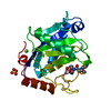
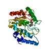
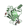
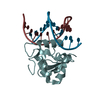
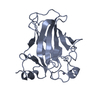
CRYSTAL STRUCTURE OF THE MOUSE P53 CORE DNA-BINDING DOMAIN AT 2.7A RESOLUTION
要素
CELLULAR TUMOR ANTIGEN P53
キーワード
DNA BINDING PROTEIN / p53 / tumor suppressor / DNA binding
機能・相同性
機能・相同性情報
Formation of Senescence-Associated Heterochromatin Foci (SAHF) / Regulation of TP53 Expression / Regulation of TP53 Activity through Acetylation / Transcriptional activation of cell cycle inhibitor p21 / Regulation of TP53 Activity through Association with Co-factors / regulation of thymocyte apoptotic process / PI5P Regulates TP53 Acetylation / RUNX3 regulates CDKN1A transcription / Stabilization of p53 / DNA Damage/Telomere Stress Induced Senescence ...Formation of Senescence-Associated Heterochromatin Foci (SAHF) / Regulation of TP53 Expression / Regulation of TP53 Activity through Acetylation / Transcriptional activation of cell cycle inhibitor p21 / Regulation of TP53 Activity through Association with Co-factors / regulation of thymocyte apoptotic process / PI5P Regulates TP53 Acetylation / RUNX3 regulates CDKN1A transcription / Stabilization of p53 / DNA Damage/Telomere Stress Induced Senescence / Regulation of TP53 Activity through Methylation / G2/M DNA damage checkpoint / Regulation of TP53 Degradation / Oncogene Induced Senescence / Autodegradation of the E3 ubiquitin ligase COP1 / G2/M Checkpoints / Ovarian tumor domain proteases / Recruitment and ATM-mediated phosphorylation of repair and signaling proteins at DNA double strand breaks / PKR-mediated signaling / The role of GTSE1 in G2/M progression after G2 checkpoint / regulation of cellular senescence / Oxidative Stress Induced Senescence / Regulation of TP53 Activity through Phosphorylation / Ub-specific processing proteases / embryo development ending in birth or egg hatching / signal transduction by p53 class mediator / negative regulation of G1 to G0 transition / negative regulation of glucose catabolic process to lactate via pyruvate / regulation of intrinsic apoptotic signaling pathway by p53 class mediator / negative regulation of pentose-phosphate shunt / ATP-dependent DNA/DNA annealing activity / regulation of cell cycle G2/M phase transition / regulation of fibroblast apoptotic process / intrinsic apoptotic signaling pathway in response to hypoxia / oligodendrocyte apoptotic process / negative regulation of miRNA processing / positive regulation of thymocyte apoptotic process / oxidative stress-induced premature senescence / regulation of tissue remodeling / negative regulation of mitotic cell cycle / positive regulation of mitochondrial membrane permeability / mRNA transcription / positive regulation of programmed necrotic cell death / bone marrow development / circadian behavior / T cell proliferation involved in immune response / regulation of mitochondrial membrane permeability involved in apoptotic process / germ cell nucleus / glucose catabolic process to lactate via pyruvate / regulation of DNA damage response, signal transduction by p53 class mediator / histone deacetylase regulator activity / negative regulation of glial cell proliferation / negative regulation of neuroblast proliferation / mitochondrial DNA repair / T cell lineage commitment / ER overload response / thymocyte apoptotic process / B cell lineage commitment / negative regulation of mitophagy / cardiac septum morphogenesis / cellular response to stress / negative regulation of DNA replication / entrainment of circadian clock by photoperiod / negative regulation of telomere maintenance via telomerase / positive regulation of release of cytochrome c from mitochondria / necroptotic process / TFIID-class transcription factor complex binding / intrinsic apoptotic signaling pathway by p53 class mediator / regulation of neuron apoptotic process / negative regulation of reactive oxygen species metabolic process / rRNA transcription / replicative senescence / cellular response to UV-C / cellular response to actinomycin D / intrinsic apoptotic signaling pathway in response to endoplasmic reticulum stress / positive regulation of execution phase of apoptosis / positive regulation of RNA polymerase II transcription preinitiation complex assembly / neuroblast proliferation / intrinsic apoptotic signaling pathway in response to DNA damage by p53 class mediator / response to X-ray / hematopoietic stem cell differentiation / viral process / embryonic organ development / chromosome organization / type II interferon-mediated signaling pathway / somitogenesis / glial cell proliferation / negative regulation of fibroblast proliferation / core promoter sequence-specific DNA binding / response to UV / positive regulation of cardiac muscle cell apoptotic process / negative regulation of stem cell proliferation / cellular response to glucose starvation / mitophagy / positive regulation of intrinsic apoptotic signaling pathway / gastrulation / response to salt stress / mitotic G1 DNA damage checkpoint signaling / negative regulation of proteolysis / MDM2/MDM4 family protein binding 類似検索 - 分子機能
 ムービー
ムービー コントローラー


 データを開く
 基本情報
基本情報 要素
要素 キーワード
キーワード 機能・相同性情報
 X線回折 /
  データ登録者
 引用
引用 構造の表示
構造の表示 ダウンロードとリンク
 その他のダウンロード
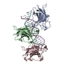


 PDBj
PDBj


 集合体
集合体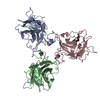


 分子量: 65.409 Da / 分子数: 3 / 由来タイプ: 合成 / 式: Zn
 分子量: 18.015 Da / 分子数: 108 / 由来タイプ: 天然 / 式: H2O
 試料調製
試料調製 / ビームライン: A1 / 波長: 1.0801 Å
 解析
解析