 Movie
Movie Controller

[English] 日本語
 Yorodumi
- PDB-1hu8: CRYSTAL STRUCTURE OF THE MOUSE P53 CORE DNA-BINDING DOMAIN AT 2.7... -
+ Open data

- Basic information
| Entry | Database: PDB / ID: 1hu8 | ||||||
|---|---|---|---|---|---|---|---|
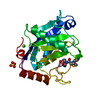
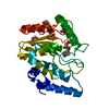
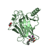
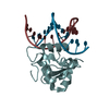
| Title | CRYSTAL STRUCTURE OF THE MOUSE P53 CORE DNA-BINDING DOMAIN AT 2.7A RESOLUTION | ||||||
 Components Components | CELLULAR TUMOR ANTIGEN P53 | ||||||
 Keywords Keywords | DNA BINDING PROTEIN / p53 / tumor suppressor / DNA binding | ||||||
| Function / homology |  Function and homology information Function and homology informationFormation of Senescence-Associated Heterochromatin Foci (SAHF) / Regulation of TP53 Expression / Regulation of TP53 Activity through Acetylation / Transcriptional activation of cell cycle inhibitor p21 / Regulation of TP53 Activity through Association with Co-factors / regulation of thymocyte apoptotic process / PI5P Regulates TP53 Acetylation / RUNX3 regulates CDKN1A transcription / DNA Damage/Telomere Stress Induced Senescence / G2/M DNA damage checkpoint ...Formation of Senescence-Associated Heterochromatin Foci (SAHF) / Regulation of TP53 Expression / Regulation of TP53 Activity through Acetylation / Transcriptional activation of cell cycle inhibitor p21 / Regulation of TP53 Activity through Association with Co-factors / regulation of thymocyte apoptotic process / PI5P Regulates TP53 Acetylation / RUNX3 regulates CDKN1A transcription / DNA Damage/Telomere Stress Induced Senescence / G2/M DNA damage checkpoint / Stabilization of p53 / Regulation of TP53 Activity through Methylation / Regulation of TP53 Degradation / negative regulation of DNA biosynthetic process / Oncogene Induced Senescence / Autodegradation of the E3 ubiquitin ligase COP1 / G2/M Checkpoints / Ovarian tumor domain proteases / Recruitment and ATM-mediated phosphorylation of repair and signaling proteins at DNA double strand breaks / PKR-mediated signaling / The role of GTSE1 in G2/M progression after G2 checkpoint / regulation of cellular senescence / Oxidative Stress Induced Senescence / Regulation of TP53 Activity through Phosphorylation / negative regulation of mitotic cell cycle / Ub-specific processing proteases / embryo development ending in birth or egg hatching / positive regulation of leukocyte migration / signal transduction by p53 class mediator / negative regulation of G1 to G0 transition / negative regulation of glucose catabolic process to lactate via pyruvate / regulation of intrinsic apoptotic signaling pathway by p53 class mediator / negative regulation of pentose-phosphate shunt / ATP-dependent DNA/DNA annealing activity / regulation of cell cycle G2/M phase transition / oligodendrocyte apoptotic process / negative regulation of miRNA processing / intrinsic apoptotic signaling pathway in response to hypoxia / oxidative stress-induced premature senescence / regulation of tissue remodeling / positive regulation of thymocyte apoptotic process / positive regulation of mitochondrial membrane permeability / regulation of fibroblast apoptotic process / bone marrow development / cellular response to actinomycin D / circadian behavior / regulation of mitochondrial membrane permeability involved in apoptotic process / histone deacetylase regulator activity / positive regulation of programmed necrotic cell death / T cell proliferation involved in immune response / mRNA transcription / negative regulation of neuroblast proliferation / regulation of DNA damage response, signal transduction by p53 class mediator / mitochondrial DNA repair / T cell lineage commitment / ER overload response / cardiac septum morphogenesis / necroptotic process / B cell lineage commitment / cellular response to stress / entrainment of circadian clock by photoperiod / negative regulation of DNA replication / negative regulation of mitophagy / positive regulation of release of cytochrome c from mitochondria / negative regulation of telomere maintenance via telomerase / rRNA transcription / negative regulation of reactive oxygen species metabolic process / intrinsic apoptotic signaling pathway by p53 class mediator / TFIID-class transcription factor complex binding / cellular response to UV-C / replicative senescence / viral process / intrinsic apoptotic signaling pathway in response to endoplasmic reticulum stress / hematopoietic stem cell differentiation / embryonic organ development / intrinsic apoptotic signaling pathway in response to DNA damage by p53 class mediator / positive regulation of RNA polymerase II transcription preinitiation complex assembly / chromosome organization / general transcription initiation factor binding / positive regulation of execution phase of apoptosis / type II interferon-mediated signaling pathway / hematopoietic progenitor cell differentiation / response to X-ray / somitogenesis / response to UV / negative regulation of fibroblast proliferation / core promoter sequence-specific DNA binding / cis-regulatory region sequence-specific DNA binding / cellular response to glucose starvation / negative regulation of proteolysis / mitotic G1 DNA damage checkpoint signaling / positive regulation of intrinsic apoptotic signaling pathway / response to salt stress / transcription repressor complex / gastrulation / 14-3-3 protein binding / positive regulation of cardiac muscle cell apoptotic process / transforming growth factor beta receptor signaling pathway / positive regulation of cell cycle / regulation of mitotic cell cycle Similarity search - Function | ||||||
| Biological species |  | ||||||
| Method |  X-RAY DIFFRACTION / SYNCHROTRON / MOLECULAR REPLACEMENT / Resolution: 2.7 Å X-RAY DIFFRACTION / SYNCHROTRON / MOLECULAR REPLACEMENT / Resolution: 2.7 Å | ||||||
 Authors Authors | Zhao, K. / Chai, X. / Johnston, K. / Clements, A. / Marmorstein, R. | ||||||
 Citation Citation | Journal: J.Biol.Chem. / Year: 2001 Title: Crystal structure of the mouse p53 core DNA-binding domain at 2.7 A resolution. Authors: Zhao, K. / Chai, X. / Johnston, K. / Clements, A. / Marmorstein, R. | ||||||
| History |
|
- Structure visualization
| Structure viewer | Molecule: MolmilJmol/JSmol |
|---|
- Downloads & links
-Download
| PDBx/mmCIF format | 1hu8.cif.gz | 122.2 KB | Display | PDBx/mmCIF format |
|---|---|---|---|---|
| PDB format | pdb1hu8.ent.gz | 96.2 KB | Display | PDB format |
| PDBx/mmJSON format | 1hu8.json.gz | Tree view | PDBx/mmJSON format | |
| Others |  Other downloads Other downloads |
-Validation report
| Arichive directory | https://data.pdbj.org/pub/pdb/validation_reports/hu/1hu8ftp://data.pdbj.org/pub/pdb/validation_reports/hu/1hu8 | HTTPS FTP |
|---|
-Related structure data
| Related structure data | 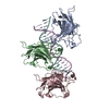 1tsrS S: Starting model for refinement |
|---|---|
| Similar structure data |


-Links
 PDBj
PDBj


- Assembly
| Deposited unit | 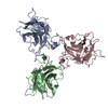
| ||||||||
|---|---|---|---|---|---|---|---|---|---|
| 1 | 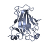
| ||||||||
| 2 | 
| ||||||||
| 3 | 
| ||||||||
| Unit cell |
|
-Components
| #1: Protein | Mass: 20928.838 Da / Num. of mol.: 3 / Fragment: RESIDUES 99-284 / Mutation: Y199A Source method: isolated from a genetically manipulated source Source: (gene. exp.)  #2: Chemical |   Mass: 65.409 Da / Num. of mol.: 3 / Source method: obtained synthetically / Formula: Zn Mass: 65.409 Da / Num. of mol.: 3 / Source method: obtained synthetically / Formula: Zn#3: Water | ChemComp-HOH / |  Mass: 18.015 Da / Num. of mol.: 108 / Source method: isolated from a natural source / Formula: H2O Mass: 18.015 Da / Num. of mol.: 108 / Source method: isolated from a natural source / Formula: H2O |
|---|
-Experimental details
-Experiment
| Experiment | Method: X-RAY DIFFRACTION / Number of used crystals: 1 |
|---|
- Sample preparation
| Crystal | Density Matthews: 3.07 Å3/Da / Density % sol: 57 % | ||||||||||||||||||||||||||||||||||||
|---|---|---|---|---|---|---|---|---|---|---|---|---|---|---|---|---|---|---|---|---|---|---|---|---|---|---|---|---|---|---|---|---|---|---|---|---|---|
| Crystal grow | Temperature: 293 K / Method: vapor diffusion, hanging drop / pH: 7.5 Details: PEG 4000, KCl and MgCl, pH 7.5, VAPOR DIFFUSION, HANGING DROP, temperature 293K | ||||||||||||||||||||||||||||||||||||
| Crystal grow | *PLUS | ||||||||||||||||||||||||||||||||||||
| Components of the solutions | *PLUS
|
-Data collection
| Diffraction | Mean temperature: 180 K |
|---|---|
| Diffraction source | Source: SYNCHROTRON / Site: CHESS  / Beamline: A1 / Wavelength: 1.0801 Å / Beamline: A1 / Wavelength: 1.0801 Å |
| Detector | Type: ADSC QUANTUM 4 / Detector: CCD / Date: Jun 7, 2000 / Details: mirrors |
| Radiation | Monochromator: YALE MIRRORS / Protocol: SINGLE WAVELENGTH / Monochromatic (M) / Laue (L): M / Scattering type: x-ray |
| Radiation wavelength | Wavelength: 1.0801 Å / Relative weight: 1 |
| Reflection | Resolution: 2.7→100 Å / Num. all: 121009 / Num. obs: 22733 / % possible obs: 99.5 % / Observed criterion σ(F): 0 / Observed criterion σ(I): 0 / Redundancy: 5.3 % / Biso Wilson estimate: 71.7 Å2 / Rmerge(I) obs: 0.051 / Net I/σ(I): 9.6 |
| Reflection shell | Resolution: 2.7→2.85 Å / Redundancy: 4.8 % / Rmerge(I) obs: 0.207 / Mean I/σ(I) obs: 3.9 / Num. unique all: 3242 / Rsym value: 0.186 / % possible all: 99.5 |
| Reflection | *PLUS Lowest resolution: 10 Å / Num. measured all: 121009 |
| Reflection shell | *PLUS % possible obs: 98.7 % / Rmerge(I) obs: 0.189 |
- Processing
| Software |
| ||||||||||||||||||||||||||||||||||||
|---|---|---|---|---|---|---|---|---|---|---|---|---|---|---|---|---|---|---|---|---|---|---|---|---|---|---|---|---|---|---|---|---|---|---|---|---|---|
| Refinement | Method to determine structure: MOLECULAR REPLACEMENT Starting model: PDB ENTRY 1TSR Resolution: 2.7→10 Å / Rfactor Rfree error: 0.006 / Data cutoff high rms absF: 1546899.25 / Isotropic thermal model: Isotropic / Cross valid method: THROUGHOUT / σ(F): 2 / σ(I): 2 / Stereochemistry target values: Engh & Huber
| ||||||||||||||||||||||||||||||||||||
| Displacement parameters | Biso mean: 63.5 Å2
| ||||||||||||||||||||||||||||||||||||
| Refine analyze |
| ||||||||||||||||||||||||||||||||||||
| Refinement step | Cycle: LAST / Resolution: 2.7→10 Å
| ||||||||||||||||||||||||||||||||||||
| Refine LS restraints |
| ||||||||||||||||||||||||||||||||||||
| LS refinement shell | Resolution: 2.7→2.79 Å / Rfactor Rfree error: 0.03 / Total num. of bins used: 10
| ||||||||||||||||||||||||||||||||||||
| Xplor file |
| ||||||||||||||||||||||||||||||||||||
| Software | *PLUS Name: CNS / Version: 0.9 / Classification: refinement | ||||||||||||||||||||||||||||||||||||
| Refinement | *PLUS Highest resolution: 2.7 Å / Lowest resolution: 10 Å / σ(F): 2 / % reflection Rfree: 9.9 % / Rfactor obs: 0.239 | ||||||||||||||||||||||||||||||||||||
| Solvent computation | *PLUS | ||||||||||||||||||||||||||||||||||||
| Displacement parameters | *PLUS | ||||||||||||||||||||||||||||||||||||
| Refine LS restraints | *PLUS
| ||||||||||||||||||||||||||||||||||||
| LS refinement shell | *PLUS Rfactor Rfree: 0.449 / Rfactor Rwork: 0.383 |
