 Movie
Movie Controller

[English] 日本語
 Yorodumi
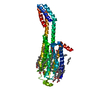
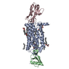
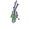
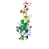
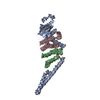
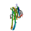
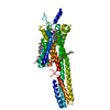
- PDB-1htm: STRUCTURE OF INFLUENZA HAEMAGGLUTININ AT THE PH OF MEMBRANE FUSION -
+ Open data

- Basic information
| Entry | Database: PDB / ID: 1htm | ||||||
|---|---|---|---|---|---|---|---|
| Title | STRUCTURE OF INFLUENZA HAEMAGGLUTININ AT THE PH OF MEMBRANE FUSION | ||||||
 Components Components |
| ||||||
 Keywords Keywords | VIRAL PROTEIN / INFLUENZA VIRUS HEMAGGLUTININ | ||||||
| Function / homology |  Function and homology information Function and homology informationviral budding from plasma membrane / clathrin-dependent endocytosis of virus by host cell / host cell surface receptor binding / fusion of virus membrane with host plasma membrane / fusion of virus membrane with host endosome membrane / viral envelope / virion attachment to host cell / host cell plasma membrane / virion membrane / membrane Similarity search - Function | ||||||
| Biological species |  uncultured beta proteobacterium UMTRA-608 (environmental samples) uncultured beta proteobacterium UMTRA-608 (environmental samples) | ||||||
| Method |  X-RAY DIFFRACTION / Resolution: 2.5 Å X-RAY DIFFRACTION / Resolution: 2.5 Å | ||||||
 Authors Authors | Bullough, P.A. / Hughson, F.M. / Skehel, J.J. / Wiley, D.C. | ||||||
 Citation Citation | Journal: Nature / Year: 1994 Title: Structure of influenza haemagglutinin at the pH of membrane fusion. Authors: Bullough, P.A. / Hughson, F.M. / Skehel, J.J. / Wiley, D.C. #1: Journal: J.Mol.Biol. / Year: 1994Title: Crystals of a Fragment of Influenza Haemagglutinin in the Low Ph Induced Conformation Authors: Bullough, P.A. / Hughson, F.M. / Treharne, A.C. / Ruigrok, R.W.H. / Skehel, J.J. / Wiley, D.C. #2: Journal: J.Mol.Biol. / Year: 1990Title: Refinement of the Influenza Virus Hemagglutinin by Simulated Annealing Authors: Weis, W.I. / Brunger, A.T. / Skehel, J.J. / Wiley, D.C. #3: Journal: J.Gen.Virol. / Year: 1988Title: Studies on the Structure of the Influenza Virus Haemagglutinin at the Ph of Membrane Fusion Authors: Ruigrok, R.W.H. / Aitken, A. / Calder, L.J. / Martin, S.R. / Skehel, J.J. / Wharton, S.A. / Weis, W. / Wiley, D.C. #4: Journal: J.Gen.Virol. / Year: 1983Title: Analyses of the Antigenicity of Influenza Haemagglutinin at the Ph Optimum for Virus-Mediated Membrane Fusion Authors: Daniels, R.S. / Douglas, A.R. / Skehel, J.J. #5: Journal: Proc.Natl.Acad.Sci.USA / Year: 1982Title: Changes in the Conformation of Influenza Virus Hemagglutinin at the Ph Optimum of Virus-Mediated Membrane Fusion Authors: Skehel, J.J. / Bayley, P.M. / Brown, E.B. / Martin, S.R. / Waterfield, M.D. / White, J.M. / Wilson, I.A. / Wiley, D.C. #6: Journal: Nature / Year: 1981Title: Structure of the Haemagglutinin Membrane Glycoprotein of Influenza Virus at 3 Angstroms Resolution Authors: Wilson, I.A. / Skehel, J.J. / Wiley, D.C. | ||||||
| History |
|
- Structure visualization
| Structure viewer | Molecule: MolmilJmol/JSmol |
|---|
- Downloads & links
-Download
| PDBx/mmCIF format | 1htm.cif.gz | 90.1 KB | Display | PDBx/mmCIF format |
|---|---|---|---|---|
| PDB format | pdb1htm.ent.gz | 69.6 KB | Display | PDB format |
| PDBx/mmJSON format | 1htm.json.gz | Tree view | PDBx/mmJSON format | |
| Others |  Other downloads Other downloads |
-Validation report
| Arichive directory | https://data.pdbj.org/pub/pdb/validation_reports/ht/1htmftp://data.pdbj.org/pub/pdb/validation_reports/ht/1htm | HTTPS FTP |
|---|
-Related structure data
| Similar structure data |
|---|

-Links
 PDBj
PDBj


- Assembly
| Deposited unit | 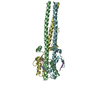
| ||||||||||||
|---|---|---|---|---|---|---|---|---|---|---|---|---|---|
| 1 |
| ||||||||||||
| Unit cell |
| ||||||||||||
| Noncrystallographic symmetry (NCS) | NCS oper:
| ||||||||||||
| Details | TBHA2 IS A TRIMER WHOSE IDENTICAL SUBUNITS CONSIST OF RESIDUES 1 - 27 OF THE HA1 CHAIN AND RESIDUES 38 - 175 OF THE HA2 CHAIN. WITHIN EACH SUBUNIT, THE HA1 AND HA2 CHAINS ARE LINKED BY A SINGLE DISULFIDE BOND BETWEEN HA1 RESIDUE 14 AND HA2 RESIDUE 137. AS IN EARLIER HA STRUCTURES (SEE REFERENCES), CHAINS HA1 AND HA2 OF SUBUNIT 1 HAVE BEEN ASSIGNED CHAIN IDENTIFIERS *A* AND *B*, RESPECTIVELY. CHAINS HA1 AND HA2 OF SUBUNIT 2 HAVE BEEN ASSIGNED CHAIN IDENTIFIERS *C* AND *D*, RESPECTIVELY. CHAINS HA1 AND HA2 OF SUBUNIT 3 HAVE BEEN ASSIGNED CHAIN IDENTIFIERS *E* AND *F*, RESPECTIVELY. IN ADDITION, 35 WATER MOLECULES/TRIMER ARE INCLUDED, NUMBERED FROM 1001 WITH NO CHAIN IDENTIFIER. THE TRANSFORMATION PRESENTED ON *MTRIX 1* RECORDS BELOW WILL YIELD APPROXIMATE COORDINATES FOR CHAINS *A* AND *B* WHEN APPLIED TO CHAINS *C* AND *D*. THE TRANSFORMATION PRESENTED ON *MTRIX 2* RECORDS BELOW WILL YIELD APPROXIMATE COORDINATES FOR CHAINS *A* AND *B* WHEN APPLIED TO CHAINS *E* AND *F*. MONOMERS 1 AND 2 SUPERIMPOSE WITH AN RMS DEVIATION BETWEEN ALPHA CARBON ATOMS OF ABOUT 0.8 ANGSTROMS; MONOMERS 1 AND 3 SUPERIMPOSE WITH AN RMS DEVIATION BETWEEN ALPHA CARBON ATOMS OF ABOUT 1.2 ANGSTROMS. |
-Components
| #1: Protein/peptide | Mass: 2776.066 Da / Num. of mol.: 3 Source method: isolated from a genetically manipulated source Source: (gene. exp.) uncultured beta proteobacterium UMTRA-608 (environmental samples)References: UniProt: P03437 #2: Protein | Mass: 16268.125 Da / Num. of mol.: 3 Source method: isolated from a genetically manipulated source Source: (gene. exp.) uncultured beta proteobacterium UMTRA-608 (environmental samples)References: UniProt: P03437 #3: Water | ChemComp-HOH / |  Mass: 18.015 Da / Num. of mol.: 37 / Source method: isolated from a natural source / Formula: H2O Mass: 18.015 Da / Num. of mol.: 37 / Source method: isolated from a natural source / Formula: H2OCompound details | IN THE VIRUS, HEMAGGLUTININ IS A TRIMER OF IDENTICAL SUBUNITS, EACH CONSISTING OF A DISULFIDE- ...IN THE VIRUS, HEMAGGLUTI | Has protein modification | Y | |
|---|
-Experimental details
-Experiment
| Experiment | Method: X-RAY DIFFRACTION |
|---|
- Sample preparation
| Crystal | Density Matthews: 4.6 Å3/Da / Density % sol: 73.26 % | ||||||||||||||||||||||||||||||||||||||||||||||||||||||
|---|---|---|---|---|---|---|---|---|---|---|---|---|---|---|---|---|---|---|---|---|---|---|---|---|---|---|---|---|---|---|---|---|---|---|---|---|---|---|---|---|---|---|---|---|---|---|---|---|---|---|---|---|---|---|---|
| Crystal grow | *PLUS pH: 5 / Method: vapor diffusion, hanging drop / Details: Bullough, P.A., (1994) J.Mol.Biol., 236, 1262. | ||||||||||||||||||||||||||||||||||||||||||||||||||||||
| Components of the solutions | *PLUS
|
-Data collection
| Radiation | Scattering type: x-ray |
|---|---|
| Radiation wavelength | Relative weight: 1 |
| Reflection | Resolution: 2.5→6 Å / Num. obs: 27793 / % possible obs: 81.05 % / Observed criterion σ(I): 2 |
- Processing
| Software |
| ||||||||||||||||||||||||||||||||||||||||||||||||||||||||||||
|---|---|---|---|---|---|---|---|---|---|---|---|---|---|---|---|---|---|---|---|---|---|---|---|---|---|---|---|---|---|---|---|---|---|---|---|---|---|---|---|---|---|---|---|---|---|---|---|---|---|---|---|---|---|---|---|---|---|---|---|---|---|
| Refinement | Rfactor Rfree: 0.291 / Rfactor Rwork: 0.222 / Rfactor obs: 0.222 / Highest resolution: 2.5 Å | ||||||||||||||||||||||||||||||||||||||||||||||||||||||||||||
| Refinement step | Cycle: LAST / Highest resolution: 2.5 Å
| ||||||||||||||||||||||||||||||||||||||||||||||||||||||||||||
| Refine LS restraints |
| ||||||||||||||||||||||||||||||||||||||||||||||||||||||||||||
| Software | *PLUS Name: X-PLOR / Classification: refinement | ||||||||||||||||||||||||||||||||||||||||||||||||||||||||||||
| Refinement | *PLUS Highest resolution: 2.5 Å / Lowest resolution: 6 Å / Num. reflection obs: 25888 / Rfactor obs: 0.222 / Rfactor Rfree: 0.291 / Rfactor Rwork: 0.222 | ||||||||||||||||||||||||||||||||||||||||||||||||||||||||||||
| Solvent computation | *PLUS | ||||||||||||||||||||||||||||||||||||||||||||||||||||||||||||
| Displacement parameters | *PLUS | ||||||||||||||||||||||||||||||||||||||||||||||||||||||||||||
| Refine LS restraints | *PLUS Type: x_angle_d / Dev ideal: 2.17 |
