 Movie
Movie Controller

[English] 日本語
 Yorodumi
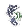
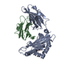
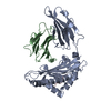
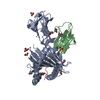
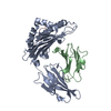
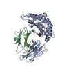
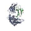
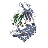
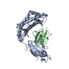
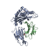
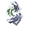
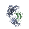
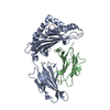
- PDB-1hsa: THE THREE-DIMENSIONAL STRUCTURE OF HLA-B27 AT 2.1 ANGSTROMS RESOL... -
+ Open data

- Basic information
| Entry | Database: PDB / ID: 1hsa | ||||||
|---|---|---|---|---|---|---|---|
| Title | THE THREE-DIMENSIONAL STRUCTURE OF HLA-B27 AT 2.1 ANGSTROMS RESOLUTION SUGGESTS A GENERAL MECHANISM FOR TIGHT PEPTIDE BINDING TO MHC | ||||||
 Components Components |
| ||||||
 Keywords Keywords | HISTOCOMPATIBILITY ANTIGEN | ||||||
| Function / homology |  Function and homology information Function and homology informationregulation of interleukin-12 production / regulation of dendritic cell differentiation / regulation of T cell anergy / regulation of interleukin-6 production / protection from natural killer cell mediated cytotoxicity / TAP binding / detection of bacterium / antigen processing and presentation of endogenous peptide antigen via MHC class Ib / antigen processing and presentation of endogenous peptide antigen via MHC class I via ER pathway, TAP-independent / early endosome lumen ...regulation of interleukin-12 production / regulation of dendritic cell differentiation / regulation of T cell anergy / regulation of interleukin-6 production / protection from natural killer cell mediated cytotoxicity / TAP binding / detection of bacterium / antigen processing and presentation of endogenous peptide antigen via MHC class Ib / antigen processing and presentation of endogenous peptide antigen via MHC class I via ER pathway, TAP-independent / early endosome lumen / Nef mediated downregulation of MHC class I complex cell surface expression / DAP12 interactions / secretory granule membrane / T cell mediated cytotoxicity / Endosomal/Vacuolar pathway / Antigen Presentation: Folding, assembly and peptide loading of class I MHC / lumenal side of endoplasmic reticulum membrane / regulation of iron ion transport / cellular response to iron(III) ion / antigen processing and presentation of exogenous protein antigen via MHC class Ib, TAP-dependent / negative regulation of iron ion transport / negative regulation of forebrain neuron differentiation / regulation of erythrocyte differentiation / peptide antigen assembly with MHC class I protein complex / ER to Golgi transport vesicle membrane / response to molecule of bacterial origin / defense response / HFE-transferrin receptor complex / MHC class I peptide loading complex / transferrin transport / negative regulation of receptor-mediated endocytosis / cellular response to iron ion / positive regulation of T cell cytokine production / antigen processing and presentation of endogenous peptide antigen via MHC class I / MHC class I protein complex / peptide antigen assembly with MHC class II protein complex / negative regulation of neurogenesis / multicellular organismal-level iron ion homeostasis / cellular response to nicotine / MHC class II protein complex / positive regulation of receptor-mediated endocytosis / positive regulation of T cell mediated cytotoxicity / negative regulation of epithelial cell proliferation / specific granule lumen / antigen processing and presentation of exogenous peptide antigen via MHC class II / positive regulation of immune response / peptide antigen binding / phagocytic vesicle membrane / recycling endosome membrane / positive regulation of T cell activation / Interferon gamma signaling / Immunoregulatory interactions between a Lymphoid and a non-Lymphoid cell / Interferon alpha/beta signaling / Modulation by Mtb of host immune system / sensory perception of smell / positive regulation of cellular senescence / tertiary granule lumen / MHC class II protein complex binding / T cell differentiation in thymus / DAP12 signaling / late endosome membrane / negative regulation of neuron projection development / protein refolding / protein-folding chaperone binding / ER-Phagosome pathway / early endosome membrane / amyloid fibril formation / protein homotetramerization / intracellular iron ion homeostasis / adaptive immune response / learning or memory / immune response / endoplasmic reticulum lumen / Amyloid fiber formation / Golgi membrane / external side of plasma membrane / signaling receptor binding / innate immune response / lysosomal membrane / focal adhesion / Neutrophil degranulation / SARS-CoV-2 activates/modulates innate and adaptive immune responses / structural molecule activity / Golgi apparatus / cell surface / endoplasmic reticulum / protein homodimerization activity / : / extracellular exosome / extracellular region / membrane / identical protein binding / plasma membrane / cytosol Similarity search - Function | ||||||
| Biological species |  Homo sapiens (human) Homo sapiens (human) | ||||||
| Method |  X-RAY DIFFRACTION / Resolution: 2.1 Å X-RAY DIFFRACTION / Resolution: 2.1 Å | ||||||
 Authors Authors | Madden, D.R. / Gorga, J.C. / Strominger, J.L. / Wiley, D.C. | ||||||
 Citation Citation | Journal: Cell(Cambridge,Mass.) / Year: 1992 Title: The three-dimensional structure of HLA-B27 at 2.1 A resolution suggests a general mechanism for tight peptide binding to MHC. Authors: Madden, D.R. / Gorga, J.C. / Strominger, J.L. / Wiley, D.C. #1: Journal: Nature / Year: 1991Title: The Structure of Hla-B27 Reveals Nonamer Self-Peptides Bound in an Extended Conformation Authors: Madden, D.R. / Gorga, J.C. / Strominger, J.L. / Wiley, D.C. #2: Journal: Nature / Year: 1991Title: Identification of Self Peptides Bound to Purified Hla-B27 Authors: Jardetzky, T.S. / Lane, W.S. / Robinson, R.A. / Madden, D.R. / Wiley, D.C. #3: Journal: Proteins / Year: 1992Title: Crystallization and Preliminary X-Ray Diffraction Studies of the Human Major Histocompatibility Antigen Hla-B27 Authors: Gorga, J.C. / Madden, D.R. / Prendergast, J.K. / Wiley, D.C. / Strominger, J.L. #4: Journal: J.Mol.Biol. / Year: 1991Title: Refined Structure of the Human Histocompatibility Antigen Hla-A2 at 2.6 Angstroms Resolution Authors: Saper, M.A. / Bjorkman, P.J. / Wiley, D.C. #5: Journal: Nature / Year: 1989Title: Specificity Pockets for the Side Chains of Peptide Antigens in Hla-Aw68 Authors: Garrett, T.P.J. / Saper, M.A. / Bjorkman, P.J. / Strominger, J.L. / Wiley, D.C. #6: Journal: Nature / Year: 1987Title: Structure of the Human Class I Histocompatibility Antigen, Hla-A2 Authors: Bjorkman, P.J. / Saper, M.A. / Samraoui, B. / Bennett, W.S. / Strominger, J.L. / Wiley, D.C. #7: Journal: Nature / Year: 1987Title: The Foreign Antigen Binding Site and T Cell Recognition Regions of Class I Histocompatibility Antigens Authors: Bjorkman, P.J. / Saper, M.A. / Samraoui, B. / Bennett, W.S. / Strominger, J.L. / Wiley, D.C. #8: Journal: J.Mol.Biol. / Year: 1985Title: Crystallization and X-Ray Diffraction Studies on the Histocompatibility Antigens Hla-A2 and Hla-A28 from Human Cell Membranes Authors: Bjorkman, P.J. / Strominger, J.L. / Wiley, D.C. | ||||||
| History |
| ||||||
| Remark 700 | SHEET SHEETS 2 AND 4 EACH HAVE ONE STRAND THAT IS BIFURCATED. THIS IS REPRESENTED BY PRESENTING THE ...SHEET SHEETS 2 AND 4 EACH HAVE ONE STRAND THAT IS BIFURCATED. THIS IS REPRESENTED BY PRESENTING THE SHEETS TWICE (DESIGNATED SHEETS SB1, SB2 AND SD1, SD2 RESPECTIVELY) WHERE THE TWO REPRESENTATIONS DIFFER IN THEIR LAST STRAND. |
- Structure visualization
| Structure viewer | Molecule: MolmilJmol/JSmol |
|---|
- Downloads & links
-Download
| PDBx/mmCIF format | 1hsa.cif.gz | 174.6 KB | Display | PDBx/mmCIF format |
|---|---|---|---|---|
| PDB format | pdb1hsa.ent.gz | 137.2 KB | Display | PDB format |
| PDBx/mmJSON format | 1hsa.json.gz | Tree view | PDBx/mmJSON format | |
| Others |  Other downloads Other downloads |
-Validation report
| Arichive directory | https://data.pdbj.org/pub/pdb/validation_reports/hs/1hsaftp://data.pdbj.org/pub/pdb/validation_reports/hs/1hsa | HTTPS FTP |
|---|
-Related structure data
| Similar structure data |
|---|


-Links
 PDBj
PDBj

- Assembly
| Deposited unit | 
| ||||||||
|---|---|---|---|---|---|---|---|---|---|
| 1 | 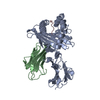
| ||||||||
| 2 | 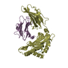
| ||||||||
| Unit cell |
| ||||||||
| Atom site foot note | 1: RESIDUES PRO A 210, PRO B 32, PRO D 210 AND PRO E 32 ARE CIS PROLINES. 2: SIDE CHAIN ATOMS OF ARG A 108, LYS A 268, LYS B 58 AND ARG D 108, LYS D 268, LYS E 58 ARE DISORDERED, AND HAVE OCCUPANCIES EQUAL TO ZERO IN THIS ENTRY. 3: THESE SOLVENT MOLECULES ARE LOCATED WITHIN THE PEPTIDE-BINDING SITE OF COMPLEX 1. 4: THESE SOLVENT MOLECULES ARE LOCATED WITHIN THE PEPTIDE-BINDING SITE OF COMPLEX 2. |
-Components
| #1: Protein | Mass: 31928.160 Da / Num. of mol.: 2 Source method: isolated from a genetically manipulated source Source: (gene. exp.) Homo sapiens (human) / References: UniProt: P03989, UniProt: P01889*PLUS#2: Protein | Mass: 11748.160 Da / Num. of mol.: 2 Source method: isolated from a genetically manipulated source Source: (gene. exp.) Homo sapiens (human) / References: UniProt: P61769#3: Protein/peptide | Mass: 743.831 Da / Num. of mol.: 2 Source method: isolated from a genetically manipulated source #4: Water | ChemComp-HOH / |  Mass: 18.015 Da / Num. of mol.: 440 / Source method: isolated from a natural source / Formula: H2O Mass: 18.015 Da / Num. of mol.: 440 / Source method: isolated from a natural source / Formula: H2OCompound details | SECONDARY STRUCTURE SPECIFICATIONS WERE MADE BY USE OF THE PROCEDURE OF W. KABSCH AND C. SANDER ...SECONDARY STRUCTURE SPECIFICAT | Has protein modification | Y | |
|---|
-Experimental details
-Experiment
| Experiment | Method: X-RAY DIFFRACTION |
|---|
- Sample preparation
| Crystal | Density Matthews: 2.83 Å3/Da / Density % sol: 56.54 % | ||||||||||||||||||||||||||||||
|---|---|---|---|---|---|---|---|---|---|---|---|---|---|---|---|---|---|---|---|---|---|---|---|---|---|---|---|---|---|---|---|
| Crystal grow | Details: THE FRAGMENT CRYSTALLIZED WAS THE EXTRACELLULAR PORTION OF THE PROTEIN CLEAVED FROM DETERGENT MICELLES WITH PAPAIN | ||||||||||||||||||||||||||||||
| Crystal grow | *PLUS Method: vapor diffusion, hanging drop / pH: 8.5 | ||||||||||||||||||||||||||||||
| Components of the solutions | *PLUS
|
-Data collection
| Radiation | Scattering type: x-ray |
|---|---|
| Radiation wavelength | Relative weight: 1 |
| Reflection | *PLUS Highest resolution: 2.1 Å / Lowest resolution: 10 Å / % possible obs: 92 % / Rmerge(I) obs: 0.077 |
- Processing
| Software |
| ||||||||||||||||||||||||||||||||||||||||||||||||||||||||||||
|---|---|---|---|---|---|---|---|---|---|---|---|---|---|---|---|---|---|---|---|---|---|---|---|---|---|---|---|---|---|---|---|---|---|---|---|---|---|---|---|---|---|---|---|---|---|---|---|---|---|---|---|---|---|---|---|---|---|---|---|---|---|
| Refinement | Rfactor Rwork: 0.203 / Rfactor obs: 0.203 / Highest resolution: 2.1 Å | ||||||||||||||||||||||||||||||||||||||||||||||||||||||||||||
| Refinement step | Cycle: LAST / Highest resolution: 2.1 Å
| ||||||||||||||||||||||||||||||||||||||||||||||||||||||||||||
| Refine LS restraints |
| ||||||||||||||||||||||||||||||||||||||||||||||||||||||||||||
| Software | *PLUS Name: X-PLOR / Classification: refinement | ||||||||||||||||||||||||||||||||||||||||||||||||||||||||||||
| Refinement | *PLUS Lowest resolution: 5.5 Å / Rfactor obs: 0.203 / Rfactor Rfree: 0.267 / Rfactor Rwork: 0.203 | ||||||||||||||||||||||||||||||||||||||||||||||||||||||||||||
| Solvent computation | *PLUS | ||||||||||||||||||||||||||||||||||||||||||||||||||||||||||||
| Displacement parameters | *PLUS Biso mean: 23.9 Å2 | ||||||||||||||||||||||||||||||||||||||||||||||||||||||||||||
| Refine LS restraints | *PLUS
|
