 Movie
Movie Controller

[English] 日本語
 Yorodumi
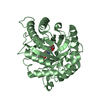
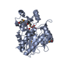
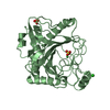
- PDB-1hn4: PROPHOSPHOLIPASE A2 DIMER COMPLEXED WITH MJ33, SULFATE, AND CALCIUM -
+ Open data

- Basic information
| Entry | Database: PDB / ID: 1hn4 | ||||||
|---|---|---|---|---|---|---|---|
| Title | PROPHOSPHOLIPASE A2 DIMER COMPLEXED WITH MJ33, SULFATE, AND CALCIUM | ||||||
 Components Components | PROPHOSPHOLIPASE A2 | ||||||
 Keywords Keywords | HYDROLASE / Enzyme / Carboxylic Ester Hydrolase / Dimer / Sulfate Binding / Inhibitor Binding | ||||||
| Function / homology |  Function and homology information Function and homology informationregulation of D-glucose import across plasma membrane / positive regulation of podocyte apoptotic process / phosphatidylglycerol metabolic process / phosphatidylcholine metabolic process / A2-type glycerophospholipase activity / leukotriene biosynthetic process / : / bile acid binding / phospholipase A2 / neutrophil mediated immunity ...regulation of D-glucose import across plasma membrane / positive regulation of podocyte apoptotic process / phosphatidylglycerol metabolic process / phosphatidylcholine metabolic process / A2-type glycerophospholipase activity / leukotriene biosynthetic process / : / bile acid binding / phospholipase A2 / neutrophil mediated immunity / positive regulation of calcium ion transport into cytosol / lipid catabolic process / neutrophil chemotaxis / positive regulation of interleukin-8 production / phospholipid binding / positive regulation of immune response / positive regulation of fibroblast proliferation / fatty acid biosynthetic process / cellular response to insulin stimulus / positive regulation of MAPK cascade / intracellular signal transduction / signaling receptor binding / calcium ion binding / positive regulation of cell population proliferation / cell surface / positive regulation of transcription by RNA polymerase II / extracellular region Similarity search - Function | ||||||
| Biological species |  | ||||||
| Method |  X-RAY DIFFRACTION / MOLECULAR REPLACEMENT / Resolution: 1.5 Å X-RAY DIFFRACTION / MOLECULAR REPLACEMENT / Resolution: 1.5 Å | ||||||
 Authors Authors | Epstein, T.M. / Pan, Y.H. / Jain, M.K. / Bahnson, B.J. | ||||||
 Citation Citation | Journal: Biochemistry / Year: 2001 Title: The basis for k(cat) impairment in prophospholipase A(2) from the anion-assisted dimer structure. Authors: Epstein, T.M. / Yu, B.Z. / Pan, Y.H. / Tutton, S.P. / Maliwal, B.P. / Jain, M.K. / Bahnson, B.J. | ||||||
| History |
|
- Structure visualization
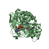
| Structure viewer | Molecule: MolmilJmol/JSmol |
|---|
- Downloads & links
-Download
| PDBx/mmCIF format | 1hn4.cif.gz | 72.4 KB | Display | PDBx/mmCIF format |
|---|---|---|---|---|
| PDB format | pdb1hn4.ent.gz | 52.5 KB | Display | PDB format |
| PDBx/mmJSON format | 1hn4.json.gz | Tree view | PDBx/mmJSON format | |
| Others |  Other downloads Other downloads |
-Validation report
| Arichive directory | https://data.pdbj.org/pub/pdb/validation_reports/hn/1hn4ftp://data.pdbj.org/pub/pdb/validation_reports/hn/1hn4 | HTTPS FTP |
|---|
-Related structure data
| Related structure data | 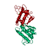 1p2pS S: Starting model for refinement |
|---|---|
| Similar structure data |


-Links
 PDBj
PDBj

- Assembly
| Deposited unit | 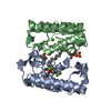
| ||||||||
|---|---|---|---|---|---|---|---|---|---|
| 1 |
| ||||||||
| Unit cell |
|
-Components
| #1: Protein | Mass: 14768.517 Da / Num. of mol.: 2 Source method: isolated from a genetically manipulated source Source: (gene. exp.)  #2: Chemical |   Mass: 40.078 Da / Num. of mol.: 2 / Source method: obtained synthetically / Formula: Ca Mass: 40.078 Da / Num. of mol.: 2 / Source method: obtained synthetically / Formula: Ca#3: Chemical | ChemComp-SO4 /   Mass: 96.063 Da / Num. of mol.: 5 / Source method: obtained synthetically / Formula: SO4 Mass: 96.063 Da / Num. of mol.: 5 / Source method: obtained synthetically / Formula: SO4#4: Chemical | ChemComp-MJI / |   Mass: 492.550 Da / Num. of mol.: 1 / Source method: obtained synthetically / Formula: C22H44F3O6P Mass: 492.550 Da / Num. of mol.: 1 / Source method: obtained synthetically / Formula: C22H44F3O6P#5: Water | ChemComp-HOH / |  Mass: 18.015 Da / Num. of mol.: 294 / Source method: isolated from a natural source / Formula: H2O Mass: 18.015 Da / Num. of mol.: 294 / Source method: isolated from a natural source / Formula: H2OHas protein modification | Y | |
|---|
-Experimental details
-Experiment
| Experiment | Method: X-RAY DIFFRACTION / Number of used crystals: 1 |
|---|
- Sample preparation
| Crystal | Density Matthews: 1.92 Å3/Da / Density % sol: 35.95 % | |||||||||||||||||||||||||||||||||||||||||||||||||
|---|---|---|---|---|---|---|---|---|---|---|---|---|---|---|---|---|---|---|---|---|---|---|---|---|---|---|---|---|---|---|---|---|---|---|---|---|---|---|---|---|---|---|---|---|---|---|---|---|---|---|
| Crystal grow | Temperature: 298 K / Method: vapor diffusion, hanging drop / pH: 4.6 Details: 25% PEG 3350, 0.2 M ammonium sulfate, 0.1 M sodium acetate trihydrate, pH 4.6, VAPOR DIFFUSION, HANGING DROP, temperature 298K | |||||||||||||||||||||||||||||||||||||||||||||||||
| Crystal grow | *PLUS Temperature: 25 ℃ | |||||||||||||||||||||||||||||||||||||||||||||||||
| Components of the solutions | *PLUS
|
-Data collection
| Diffraction | Mean temperature: 93 K |
|---|---|
| Diffraction source | Source: ROTATING ANODE / Type: RIGAKU RUH3R / Wavelength: 1.5418 Å |
| Detector | Type: RIGAKU RAXIS IV / Detector: IMAGE PLATE / Date: May 10, 2000 |
| Radiation | Monochromator: Osmic Multilayer Optics / Protocol: SINGLE WAVELENGTH / Monochromatic (M) / Laue (L): M / Scattering type: x-ray |
| Radiation wavelength | Wavelength: 1.5418 Å / Relative weight: 1 |
| Reflection | Resolution: 1.5→20 Å / Num. all: 32685 / Num. obs: 32685 / % possible obs: 91 % / Observed criterion σ(F): 0 / Observed criterion σ(I): 0 / Redundancy: 7.2 % / Biso Wilson estimate: 20.2 Å2 / Rmerge(I) obs: 0.03 / Net I/σ(I): 40.6 |
| Reflection shell | Resolution: 1.5→1.56 Å / Redundancy: 2.9 % / Rmerge(I) obs: 0.191 / % possible all: 86.3 |
| Reflection | *PLUS % possible obs: 91 % / Rmerge(I) obs: 0.03 |
- Processing
| Software |
| ||||||||||||||||||||
|---|---|---|---|---|---|---|---|---|---|---|---|---|---|---|---|---|---|---|---|---|---|
| Refinement | Method to determine structure: MOLECULAR REPLACEMENT Starting model: PDB Code 1P2P Phospholipase A2 Resolution: 1.5→20 Å / σ(F): 0 / σ(I): 0 / Stereochemistry target values: Engh & Huber
| ||||||||||||||||||||
| Refinement step | Cycle: LAST / Resolution: 1.5→20 Å
| ||||||||||||||||||||
| Refine LS restraints |
| ||||||||||||||||||||
| Software | *PLUS Name: CNS / Classification: refinement | ||||||||||||||||||||
| Refinement | *PLUS Highest resolution: 1.5 Å / Lowest resolution: 20 Å / σ(F): 0 / % reflection Rfree: 10 % / Rfactor obs: 0.215 | ||||||||||||||||||||
| Solvent computation | *PLUS | ||||||||||||||||||||
| Displacement parameters | *PLUS | ||||||||||||||||||||
| Refine LS restraints | *PLUS Type: c_angle_deg / Dev ideal: 1.1 |
