 Movie
Movie Controller

[English] 日本語
 Yorodumi
- PDB-1hl7: Gamma lactamase from an Aureobacterium species in complex with 3a... -
+ Open data

- Basic information
| Entry | Database: PDB / ID: 1hl7 | ||||||
|---|---|---|---|---|---|---|---|
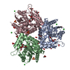
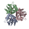
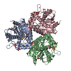
| Title | Gamma lactamase from an Aureobacterium species in complex with 3a,4,7,7a-tetrahydro-benzo [1,3] dioxol-2-one | ||||||
 Components Components | GAMMA LACTAMASE | ||||||
 Keywords Keywords | HYDROLASE / ALPHA/BETA HYDROLASE / CO-FACTOR FREE HALOPEROXIDASE / TETRAHEDRAL INTERMEDIATE | ||||||
| Function / homology |  Function and homology information Function and homology information | ||||||
| Biological species |  MICROBACTERIUM SP. (bacteria) MICROBACTERIUM SP. (bacteria) | ||||||
| Method |  X-RAY DIFFRACTION / SYNCHROTRON / MOLECULAR REPLACEMENT / Resolution: 1.73 Å X-RAY DIFFRACTION / SYNCHROTRON / MOLECULAR REPLACEMENT / Resolution: 1.73 Å | ||||||
 Authors Authors | Line, K. / Isupov, M.N. / Littlechild, J.A. | ||||||
 Citation Citation | Journal: J.Mol.Biol. / Year: 2004 Title: The Crystal Structure of a (-)Gamma-Lactamase from an Aureobacterium Species Reveals a Tetrahedral Intermediate in the Active Site Authors: Line, K. / Isupov, M.N. / Littlechild, J.A. | ||||||
| History |
|
- Structure visualization
| Structure viewer | Molecule: MolmilJmol/JSmol |
|---|
- Downloads & links
-Download
| PDBx/mmCIF format | 1hl7.cif.gz | 133.6 KB | Display | PDBx/mmCIF format |
|---|---|---|---|---|
| PDB format | pdb1hl7.ent.gz | 106.9 KB | Display | PDB format |
| PDBx/mmJSON format | 1hl7.json.gz | Tree view | PDBx/mmJSON format | |
| Others |  Other downloads Other downloads |
-Validation report
| Arichive directory | https://data.pdbj.org/pub/pdb/validation_reports/hl/1hl7ftp://data.pdbj.org/pub/pdb/validation_reports/hl/1hl7 | HTTPS FTP |
|---|
-Related structure data
| Related structure data |  1hkhC 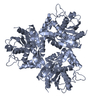 1broS C: citing same article ( S: Starting model for refinement |
|---|---|
| Similar structure data |


-Links
 PDBj
PDBj


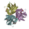
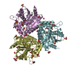
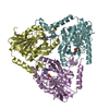
- Assembly
| Deposited unit | 
| ||||||||
|---|---|---|---|---|---|---|---|---|---|
| 1 | 
| ||||||||
| 2 | 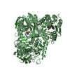
| ||||||||
| Unit cell |
| ||||||||
| Noncrystallographic symmetry (NCS) | NCS oper: (Code: given Matrix: (0.32744, 0.77849, -0.53548), Vector: |
-Components
| #1: Protein | Mass: 30878.264 Da / Num. of mol.: 2 Source method: isolated from a genetically manipulated source Source: (gene. exp.) MICROBACTERIUM SP. (bacteria) / Description: CHIROTECH CULTURE COLLECTION / Plasmid: PTRC99 / Production host: #2: Chemical | 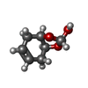  Mass: 142.152 Da / Num. of mol.: 2 / Source method: obtained synthetically / Formula: C7H10O3 Mass: 142.152 Da / Num. of mol.: 2 / Source method: obtained synthetically / Formula: C7H10O3#3: Water | ChemComp-HOH / |  Mass: 18.015 Da / Num. of mol.: 681 / Source method: isolated from a natural source / Formula: H2O Mass: 18.015 Da / Num. of mol.: 681 / Source method: isolated from a natural source / Formula: H2OHas protein modification | Y | Sequence details | NAMED CO-FACTOR FREE HALOPEROXI | |
|---|
-Experimental details
-Experiment
| Experiment | Method: X-RAY DIFFRACTION / Number of used crystals: 1 |
|---|
- Sample preparation
| Crystal | Density Matthews: 4.72 Å3/Da / Density % sol: 73.73 % |
|---|---|
| Crystal grow | pH: 8 / Details: pH 8.00 |
-Data collection
| Diffraction | Mean temperature: 100 K |
|---|---|
| Diffraction source | Source: SYNCHROTRON / Site: EMBL/DESY, HAMBURG  / Beamline: X11 / Wavelength: 0.8482 / Beamline: X11 / Wavelength: 0.8482 |
| Detector | Type: MARRESEARCH / Detector: CCD / Date: Jun 15, 2001 |
| Radiation | Protocol: SINGLE WAVELENGTH / Monochromatic (M) / Laue (L): M / Scattering type: x-ray |
| Radiation wavelength | Wavelength: 0.8482 Å / Relative weight: 1 |
| Reflection | Resolution: 1.73→11 Å / Num. obs: 118797 / % possible obs: 100 % / Redundancy: 7.5 % / Biso Wilson estimate: 15.9 Å2 / Rmerge(I) obs: 0.079 / Net I/σ(I): 28.18 |
| Reflection shell | Resolution: 1.73→1.76 Å / Redundancy: 7.2 % / Rmerge(I) obs: 0.345 / Mean I/σ(I) obs: 4.5 / % possible all: 100 |
- Processing
| Software |
| ||||||||||||||||||||
|---|---|---|---|---|---|---|---|---|---|---|---|---|---|---|---|---|---|---|---|---|---|
| Refinement | Method to determine structure: MOLECULAR REPLACEMENT Starting model: PDB ENTRY 1BRO Resolution: 1.73→11 Å / SU B: 0.981 / SU ML: 0.033 / Cross valid method: THROUGHOUT / ESU R: 0.061 / ESU R Free: 0.06
| ||||||||||||||||||||
| Displacement parameters | Biso mean: 15.6 Å2 | ||||||||||||||||||||
| Refinement step | Cycle: LAST / Resolution: 1.73→11 Å
|
