: / HSF1-dependent transactivation / Interferon gamma signaling / peptidyl-threonine autophosphorylation / Ion transport by P-type ATPases / RAF activation / calcium- and calmodulin-dependent protein kinase complex / regulation of endocannabinoid signaling pathway / neurotransmitter receptor transport to plasma membrane / Ca2+ pathway ...: / HSF1-dependent transactivation / Interferon gamma signaling / peptidyl-threonine autophosphorylation / Ion transport by P-type ATPases / RAF activation / calcium- and calmodulin-dependent protein kinase complex / regulation of endocannabinoid signaling pathway / neurotransmitter receptor transport to plasma membrane / Ca2+ pathway / Unblocking of NMDA receptors, glutamate binding and activation / Ca2+/calmodulin-dependent protein kinase / Trafficking of AMPA receptors / RAF/MAP kinase cascade / dendritic spine development / negative regulation of hydrolase activity / regulation of neurotransmitter secretion / Ion homeostasis / positive regulation of calcium ion transport / regulation of neuron migration / calcium/calmodulin-dependent protein kinase activity / regulation of mitochondrial membrane permeability involved in apoptotic process / GTPase activating protein binding / dendrite morphogenesis / regulation of neurotransmitter receptor localization to postsynaptic specialization membrane / negative regulation of ferroptosis / regulation of neuronal synaptic plasticity / glutamate receptor binding / postsynaptic cytosol / cell surface receptor signaling pathway via JAK-STAT / regulation of protein localization to plasma membrane / presynaptic cytosol / cellular response to interferon-beta / positive regulation of cardiac muscle cell apoptotic process / ionotropic glutamate receptor signaling pathway / dendrite cytoplasm / response to ischemia / angiotensin-activated signaling pathway / positive regulation of receptor signaling pathway via JAK-STAT / G1/S transition of mitotic cell cycle / cellular response to type II interferon / Schaffer collateral - CA1 synapse / kinase activity / calcium ion transport / dendritic spine / calmodulin binding / neuron projection / postsynaptic density / protein serine kinase activity / axon / protein serine/threonine kinase activity / neuronal cell body / synapse / dendrite / glutamatergic synapse / protein homodimerization activity / mitochondrion / ATP binding / metal ion binding / identical protein binding / cytoplasm Similarity search - Function
Calcium/calmodulin-dependent protein kinase II, association-domain / Calcium/calmodulin dependent protein kinase II association domain / Nuclear Transport Factor 2; Chain: A, - #50 / NTF2-like domain superfamily / Nuclear Transport Factor 2; Chain: A, / Serine/threonine-protein kinase, active site / Serine/Threonine protein kinases active-site signature. / Protein kinase domain / Serine/Threonine protein kinases, catalytic domain / Roll ...Calcium/calmodulin-dependent protein kinase II, association-domain / Calcium/calmodulin dependent protein kinase II association domain / Nuclear Transport Factor 2; Chain: A, - #50 / NTF2-like domain superfamily / Nuclear Transport Factor 2; Chain: A, / Serine/threonine-protein kinase, active site / Serine/Threonine protein kinases active-site signature. / Protein kinase domain / Serine/Threonine protein kinases, catalytic domain / Roll / Protein kinase, ATP binding site / Protein kinases ATP-binding region signature. / Protein kinase domain profile. / Protein kinase domain / Protein kinase-like domain superfamily / Alpha Beta Similarity search - Domain/homology
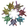
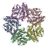
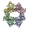
2,3-DIHYDROXY-1,4-DITHIOBUTANE / HEXATANTALUM DODECABROMIDE / Calcium/calmodulin-dependent protein kinase type II subunit alpha Similarity search - Component
Biological species
MUS MUSCULUS (house mouse)
Method
X-RAY DIFFRACTION / SYNCHROTRON / MAD / Resolution: 2.65 Å
#89 - May 2007 Aconitase and Iron Regulatory Protein 1 similarity (1)
-
Assembly
Deposited unit
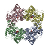
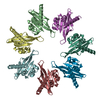
A: CALCIUM/CALMODULIN-DEPENDENT PROTEIN KINASE TYPE II ALPHA CHAIN B: CALCIUM/CALMODULIN-DEPENDENT PROTEIN KINASE TYPE II ALPHA CHAIN C: CALCIUM/CALMODULIN-DEPENDENT PROTEIN KINASE TYPE II ALPHA CHAIN D: CALCIUM/CALMODULIN-DEPENDENT PROTEIN KINASE TYPE II ALPHA CHAIN E: CALCIUM/CALMODULIN-DEPENDENT PROTEIN KINASE TYPE II ALPHA CHAIN F: CALCIUM/CALMODULIN-DEPENDENT PROTEIN KINASE TYPE II ALPHA CHAIN G: CALCIUM/CALMODULIN-DEPENDENT PROTEIN KINASE TYPE II ALPHA CHAIN H: CALCIUM/CALMODULIN-DEPENDENT PROTEIN KINASE TYPE II ALPHA CHAIN I: CALCIUM/CALMODULIN-DEPENDENT PROTEIN KINASE TYPE II ALPHA CHAIN J: CALCIUM/CALMODULIN-DEPENDENT PROTEIN KINASE TYPE II ALPHA CHAIN K: CALCIUM/CALMODULIN-DEPENDENT PROTEIN KINASE TYPE II ALPHA CHAIN L: CALCIUM/CALMODULIN-DEPENDENT PROTEIN KINASE TYPE II ALPHA CHAIN M: CALCIUM/CALMODULIN-DEPENDENT PROTEIN KINASE TYPE II ALPHA CHAIN N: CALCIUM/CALMODULIN-DEPENDENT PROTEIN KINASE TYPE II ALPHA CHAIN hetero molecules
Mass: 18.015 Da / Num. of mol.: 62 / Source method: isolated from a natural source / Formula: H2O
Compound details
THIS KINASE MAY PLAY A ROLE IN NEUROTRANSMISSION. CATALYES ATP + PROTEIN = ADP + O-PHOSPHOPROTEIN. ...THIS KINASE MAY PLAY A ROLE IN NEUROTRANSMISSION. CATALYES ATP + PROTEIN = ADP + O-PHOSPHOPROTEIN. AUTOPHOSPHORYLATION OF THR-286 MAY ALLOW THE KINASE TO SWITCH FROM A CALMODULIN-DEPENDENT TO A CALMODULIN- INDEPENDENT STATE.
-
Experimental details
-
Experiment
Experiment
Method: X-RAY DIFFRACTION / Number of used crystals: 4
In the structure databanks used in Yorodumi, some data are registered as the other names, "COVID-19 virus" and "2019-nCoV". Here are the details of the virus and the list of structure data.
Jan 31, 2019. EMDB accession codes are about to change! (news from PDBe EMDB page)
EMDB accession codes are about to change! (news from PDBe EMDB page)
The allocation of 4 digits for EMDB accession codes will soon come to an end. Whilst these codes will remain in use, new EMDB accession codes will include an additional digit and will expand incrementally as the available range of codes is exhausted. The current 4-digit format prefixed with “EMD-” (i.e. EMD-XXXX) will advance to a 5-digit format (i.e. EMD-XXXXX), and so on. It is currently estimated that the 4-digit codes will be depleted around Spring 2019, at which point the 5-digit format will come into force.
The EM Navigator/Yorodumi systems omit the EMD- prefix.
Related info.:Q: What is EMD? / ID/Accession-code notation in Yorodumi/EM Navigator
Yorodumi is a browser for structure data from EMDB, PDB, SASBDB, etc.
This page is also the successor to EM Navigator detail page, and also detail information page/front-end page for Omokage search.
The word "yorodu" (or yorozu) is an old Japanese word meaning "ten thousand". "mi" (miru) is to see.
Related info.:EMDB / PDB / SASBDB / Comparison of 3 databanks / Yorodumi Search / Aug 31, 2016. New EM Navigator & Yorodumi / Yorodumi Papers / Jmol/JSmol / Function and homology information / Changes in new EM Navigator and Yorodumi
 Movie
Movie Controller


 Open data
 Basic information
 Components
 Keywords
 Function and homology information
 X-RAY DIFFRACTION /
  Authors
 Citation
 Structure visualization
 Downloads & links
 Other downloads
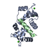

 PDBj
PDBj


 Assembly
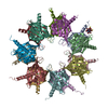


 Mass: 154.251 Da / Num. of mol.: 1 / Source method: obtained synthetically / Formula: C4H10O2S2
 Mass: 35.453 Da / Num. of mol.: 14 / Source method: obtained synthetically / Formula: Cl
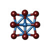
 Mass: 2044.535 Da / Num. of mol.: 1 / Source method: obtained synthetically / Formula: Br12Ta6
 Mass: 18.015 Da / Num. of mol.: 62 / Source method: isolated from a natural source / Formula: H2O
 Sample preparation
 Processing