 Movie
Movie Controller

[English] 日本語
 Yorodumi
- PDB-1cdm: MODULATION OF CALMODULIN PLASTICITY IN MOLECULAR RECOGNITION ON T... -
+ Open data

- Basic information
| Entry | Database: PDB / ID: 1cdm | ||||||
|---|---|---|---|---|---|---|---|
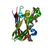
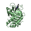
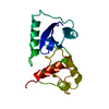
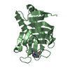
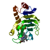
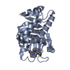
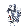
| Title | MODULATION OF CALMODULIN PLASTICITY IN MOLECULAR RECOGNITION ON THE BASIS OF X-RAY STRUCTURES | ||||||
 Components Components |
| ||||||
 Keywords Keywords | CALCIUM-BINDING PROTEIN | ||||||
| Function / homology |  Function and homology information Function and homology information: / peptidyl-threonine autophosphorylation / HSF1-dependent transactivation / neurotransmitter receptor transport to plasma membrane / RAF activation / Ion transport by P-type ATPases / calcium- and calmodulin-dependent protein kinase complex / regulation of endocannabinoid signaling pathway / Interferon gamma signaling / Ca2+/calmodulin-dependent protein kinase ...: / peptidyl-threonine autophosphorylation / HSF1-dependent transactivation / neurotransmitter receptor transport to plasma membrane / RAF activation / Ion transport by P-type ATPases / calcium- and calmodulin-dependent protein kinase complex / regulation of endocannabinoid signaling pathway / Interferon gamma signaling / Ca2+/calmodulin-dependent protein kinase / negative regulation of hydrolase activity / dendritic spine development / regulation of neuron migration / regulation of neurotransmitter secretion / Trafficking of AMPA receptors / positive regulation of calcium ion transport / calcium/calmodulin-dependent protein kinase activity / dendrite morphogenesis / Ca2+ pathway / regulation of mitochondrial membrane permeability involved in apoptotic process / RAF/MAP kinase cascade / GTPase activating protein binding / Ion homeostasis / regulation of neurotransmitter receptor localization to postsynaptic specialization membrane / calcineurin-mediated signaling / negative regulation of ferroptosis / regulation of neuronal synaptic plasticity / Unblocking of NMDA receptors, glutamate binding and activation / glutamate receptor binding / detection of calcium ion / postsynaptic cytosol / cell surface receptor signaling pathway via JAK-STAT / cellular response to interferon-beta / regulation of protein localization to plasma membrane / presynaptic cytosol / regulation of release of sequestered calcium ion into cytosol by sarcoplasmic reticulum / positive regulation of cardiac muscle cell apoptotic process / ionotropic glutamate receptor signaling pathway / dendrite cytoplasm / response to ischemia / angiotensin-activated signaling pathway / positive regulation of receptor signaling pathway via JAK-STAT / G1/S transition of mitotic cell cycle / cellular response to type II interferon / Schaffer collateral - CA1 synapse / spindle pole / kinase activity / calcium ion transport / myelin sheath / dendritic spine / calmodulin binding / neuron projection / postsynaptic density / protein domain specific binding / protein serine kinase activity / axon / protein serine/threonine kinase activity / neuronal cell body / calcium ion binding / centrosome / dendrite / synapse / glutamatergic synapse / protein homodimerization activity / protein-containing complex / mitochondrion / ATP binding / metal ion binding / identical protein binding / nucleus / cytosol / cytoplasm Similarity search - Function | ||||||
| Biological species |  | ||||||
| Method |  X-RAY DIFFRACTION / Resolution: 2 Å X-RAY DIFFRACTION / Resolution: 2 Å | ||||||
 Authors Authors | Meador, W.E. / Quiocho, F.A. | ||||||
 Citation Citation | Journal: Science / Year: 1993 Title: Modulation of calmodulin plasticity in molecular recognition on the basis of x-ray structures. Authors: Meador, W.E. / Means, A.R. / Quiocho, F.A. #1: Journal: Science / Year: 1992Title: Target Enzyme Recognition by Calmodulin: 2.4 Angstroms Structure of a Calmodulin-Peptide Complex Authors: Meador, W.E. / Means, A.R. / Quiocho, F.A. | ||||||
| History |
|
- Structure visualization
| Structure viewer | Molecule: MolmilJmol/JSmol |
|---|
- Downloads & links
-Download
| PDBx/mmCIF format | 1cdm.cif.gz | 43.8 KB | Display | PDBx/mmCIF format |
|---|---|---|---|---|
| PDB format | pdb1cdm.ent.gz | 30.2 KB | Display | PDB format |
| PDBx/mmJSON format | 1cdm.json.gz | Tree view | PDBx/mmJSON format | |
| Others |  Other downloads Other downloads |
-Validation report
| Arichive directory | https://data.pdbj.org/pub/pdb/validation_reports/cd/1cdmftp://data.pdbj.org/pub/pdb/validation_reports/cd/1cdm | HTTPS FTP |
|---|
-Related structure data
| Similar structure data |
|---|


-Links
 PDBj
PDBj


- Assembly
| Deposited unit | 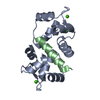
| ||||||||
|---|---|---|---|---|---|---|---|---|---|
| 1 |
| ||||||||
| Unit cell |
|
-Components
| #1: Protein | Mass: 16277.873 Da / Num. of mol.: 1 Source method: isolated from a genetically manipulated source Source: (gene. exp.) | ||
|---|---|---|---|
| #2: Protein/peptide | Mass: 2886.528 Da / Num. of mol.: 1 Source method: isolated from a genetically manipulated source References: UniProt: P11275 | ||
| #3: Chemical | ChemComp-CA /   Mass: 40.078 Da / Num. of mol.: 4 / Source method: obtained synthetically / Formula: Ca Mass: 40.078 Da / Num. of mol.: 4 / Source method: obtained synthetically / Formula: Ca#4: Water | ChemComp-HOH / |  Mass: 18.015 Da / Num. of mol.: 53 / Source method: isolated from a natural source / Formula: H2O Mass: 18.015 Da / Num. of mol.: 53 / Source method: isolated from a natural source / Formula: H2O |
-Experimental details
-Experiment
| Experiment | Method: X-RAY DIFFRACTION |
|---|
- Sample preparation
| Crystal | Density Matthews: 2.3 Å3/Da / Density % sol: 46.48 % |
|---|---|
| Crystal grow | *PLUS Method: unknown |
-Data collection
| Radiation | Scattering type: x-ray |
|---|---|
| Radiation wavelength | Relative weight: 1 |
- Processing
| Software | Name: X-PLOR / Classification: refinement | ||||||||||||||||||||||||||||||||||||||||||||||||||||||||||||
|---|---|---|---|---|---|---|---|---|---|---|---|---|---|---|---|---|---|---|---|---|---|---|---|---|---|---|---|---|---|---|---|---|---|---|---|---|---|---|---|---|---|---|---|---|---|---|---|---|---|---|---|---|---|---|---|---|---|---|---|---|---|
| Refinement | Resolution: 2→10 Å /
| ||||||||||||||||||||||||||||||||||||||||||||||||||||||||||||
| Refinement step | Cycle: LAST / Resolution: 2→10 Å
| ||||||||||||||||||||||||||||||||||||||||||||||||||||||||||||
| Refine LS restraints |
| ||||||||||||||||||||||||||||||||||||||||||||||||||||||||||||
| Software | *PLUS Name: X-PLOR / Classification: refinement | ||||||||||||||||||||||||||||||||||||||||||||||||||||||||||||
| Refinement | *PLUS Rfactor obs: 0.208 | ||||||||||||||||||||||||||||||||||||||||||||||||||||||||||||
| Solvent computation | *PLUS | ||||||||||||||||||||||||||||||||||||||||||||||||||||||||||||
| Displacement parameters | *PLUS | ||||||||||||||||||||||||||||||||||||||||||||||||||||||||||||
| Refine LS restraints | *PLUS Type: x_angle_d / Dev ideal: 1.28 |
