 Movie
Movie Controller

[English] 日本語
 Yorodumi
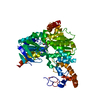
- PDB-1hfa: CALM-N N-terminal domain of clathrin assembly lymphoid myeloid le... -
+ Open data

- Basic information
| Entry | Database: PDB / ID: 1hfa | ||||||
|---|---|---|---|---|---|---|---|
| Title | CALM-N N-terminal domain of clathrin assembly lymphoid myeloid leukaemia protein, PI(4,5)P2 complex | ||||||
 Components Components | CLATHRIN ASSEMBLY PROTEIN SHORT FORM | ||||||
 Keywords Keywords | ENDOCYTOSIS / ADAPTOR | ||||||
| Function / homology |  Function and homology information Function and homology informationRND3 GTPase cycle / vesicle cargo loading / membrane bending / endosome to plasma membrane transport vesicle / regulation of terminal button organization / 1-phosphatidylinositol binding / positive regulation of synaptic vesicle clustering / regulation of protein transport / extrinsic component of presynaptic endocytic zone membrane / amyloid-beta clearance by transcytosis ...RND3 GTPase cycle / vesicle cargo loading / membrane bending / endosome to plasma membrane transport vesicle / regulation of terminal button organization / 1-phosphatidylinositol binding / positive regulation of synaptic vesicle clustering / regulation of protein transport / extrinsic component of presynaptic endocytic zone membrane / amyloid-beta clearance by transcytosis / synaptic vesicle maturation / regulation of synaptic vesicle transport / clathrin coat of coated pit / Golgi Associated Vesicle Biogenesis / negative regulation of protein localization to cell surface / regulation of vesicle size / positive regulation of amyloid precursor protein catabolic process / postsynaptic endocytic zone / clathrin heavy chain binding / positive regulation of dendrite extension / Cargo recognition for clathrin-mediated endocytosis / clathrin coat assembly / Clathrin-mediated endocytosis / positive regulation of synaptic vesicle endocytosis / vesicle budding from membrane / clathrin-dependent endocytosis / positive regulation of Ras protein signal transduction / regulation of amyloid precursor protein catabolic process / clathrin-coated vesicle / positive regulation of axonogenesis / endosomal transport / positive regulation of amyloid-beta formation / low-density lipoprotein particle receptor binding / neurofibrillary tangle / dendrite morphogenesis / clathrin binding / regulation of endocytosis / parallel fiber to Purkinje cell synapse / hemopoiesis / regulation of synaptic vesicle endocytosis / negative regulation of protein localization to plasma membrane / synaptic vesicle endocytosis / vesicle-mediated transport / phosphatidylinositol-4,5-bisphosphate binding / clathrin-coated pit / axonogenesis / receptor-mediated endocytosis / SNARE binding / negative regulation of receptor-mediated endocytosis / SH3 domain binding / multicellular organismal-level iron ion homeostasis / receptor internalization / tau protein binding / Schaffer collateral - CA1 synapse / small GTPase binding / endocytosis / synaptic vesicle / regulation of protein localization / presynaptic membrane / vesicle / intracellular iron ion homeostasis / learning or memory / early endosome / postsynaptic membrane / endosome / postsynaptic density / postsynapse / negative regulation of gene expression / neuronal cell body / positive regulation of DNA-templated transcription / perinuclear region of cytoplasm / cell surface / Golgi apparatus / membrane / identical protein binding / nucleus / plasma membrane Similarity search - Function | ||||||
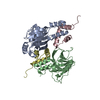
| Biological species |  | ||||||
| Method |  X-RAY DIFFRACTION / SYNCHROTRON / MOLECULAR REPLACEMENT / Resolution: 2 Å X-RAY DIFFRACTION / SYNCHROTRON / MOLECULAR REPLACEMENT / Resolution: 2 Å | ||||||
 Authors Authors | Ford, M.G.J. / Evans, P.R. / McMahon, H.T. | ||||||
 Citation Citation | Journal: Science / Year: 2001 Title: Simultaneous Binding of Ptdins(4,5)P2 and Clathrin by Ap180 in the Nucleation of Clathrin Lattices on Membranes Authors: Ford, M.G.J. / Pearse, B.M.F. / Higgins, M.K. / Vallis, Y. / Owen, D.J. / Gibson, A. / Hopkins, C.R. / Evans, P.R. / Mcmahon, H.T. | ||||||
| History |
|
- Structure visualization
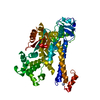
| Structure viewer | Molecule: MolmilJmol/JSmol |
|---|
- Downloads & links
-Download
| PDBx/mmCIF format | 1hfa.cif.gz | 69.7 KB | Display | PDBx/mmCIF format |
|---|---|---|---|---|
| PDB format | pdb1hfa.ent.gz | 51.6 KB | Display | PDB format |
| PDBx/mmJSON format | 1hfa.json.gz | Tree view | PDBx/mmJSON format | |
| Others |  Other downloads Other downloads |
-Validation report
| Arichive directory | https://data.pdbj.org/pub/pdb/validation_reports/hf/1hfaftp://data.pdbj.org/pub/pdb/validation_reports/hf/1hfa | HTTPS FTP |
|---|
-Related structure data
| Related structure data |  1hf8SC  1hg2C  1hg5C S: Starting model for refinement C: citing same article ( |
|---|---|
| Similar structure data |


-Links
 PDBj
PDBj


- Assembly
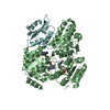
| Deposited unit | 
| ||||||||
|---|---|---|---|---|---|---|---|---|---|
| 1 | 
| ||||||||
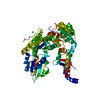
| Unit cell |
| ||||||||
| Details | BIOLOGICAL_UNIT: MONOMERTHIS DIMERIC ARRANGEMENT IS THE RESULT OF TIGHT CRYSTALPACKING. |
-Components
| #1: Protein | Mass: 32865.789 Da / Num. of mol.: 1 / Fragment: N-TERMINAL DOMAIN Source method: isolated from a genetically manipulated source Source: (gene. exp.)  |
|---|---|
| #2: Chemical | ChemComp-PIO / [(  Mass: 746.566 Da / Num. of mol.: 1 / Source method: obtained synthetically / Formula: C25H49O19P3 Mass: 746.566 Da / Num. of mol.: 1 / Source method: obtained synthetically / Formula: C25H49O19P3 |
| #3: Water | ChemComp-HOH /  Mass: 18.015 Da / Num. of mol.: 128 / Source method: isolated from a natural source / Formula: H2O Mass: 18.015 Da / Num. of mol.: 128 / Source method: isolated from a natural source / Formula: H2O |
-Experimental details
-Experiment
| Experiment | Method: X-RAY DIFFRACTION / Number of used crystals: 1 |
|---|
- Sample preparation
| Crystal | Density Matthews: 3.08 Å3/Da / Density % sol: 60 % | ||||||||||||||||||||
|---|---|---|---|---|---|---|---|---|---|---|---|---|---|---|---|---|---|---|---|---|---|
| Crystal grow | pH: 7.5 Details: 0.1M HEPES, PH 7.5, 12% PEG 8K, 8% ETHYLENE GLYCOL, CRYSTALS SOAKED IN 1MM LIGAND FOR 1 HOUR | ||||||||||||||||||||
| Crystal grow | *PLUS Method: vapor diffusion, hanging drop | ||||||||||||||||||||
| Components of the solutions | *PLUS
|
-Data collection
| Diffraction | Mean temperature: 293 K |
|---|---|
| Diffraction source | Source: SYNCHROTRON / Site: SRS  / Beamline: PX9.6 / Wavelength: 0.87 / Beamline: PX9.6 / Wavelength: 0.87 |
| Detector | Type: ADSC CCD / Detector: CCD / Date: Mar 23, 2000 |
| Radiation | Monochromator: SI(111) / Protocol: SINGLE WAVELENGTH / Monochromatic (M) / Laue (L): M / Scattering type: x-ray |
| Radiation wavelength | Wavelength: 0.87 Å / Relative weight: 1 |
| Reflection | Resolution: 2→66 Å / Num. obs: 26075 / % possible obs: 100 % / Observed criterion σ(I): 6 / Redundancy: 7.1 % / Biso Wilson estimate: 43 Å2 / Rmerge(I) obs: 0.103 / Rsym value: 0.104 / Net I/σ(I): 14.3 |
| Reflection shell | Resolution: 2→2.11 Å / Redundancy: 7 % / Rmerge(I) obs: 1.153 / Mean I/σ(I) obs: 1.5 / Rsym value: 1.153 / % possible all: 100 |
| Reflection | *PLUS % possible obs: 100 % |
| Reflection shell | *PLUS % possible obs: 100 % |
- Processing
| Software |
| ||||||||||||||||||||||||||||||||||||||||
|---|---|---|---|---|---|---|---|---|---|---|---|---|---|---|---|---|---|---|---|---|---|---|---|---|---|---|---|---|---|---|---|---|---|---|---|---|---|---|---|---|---|
| Refinement | Method to determine structure: MOLECULAR REPLACEMENT Starting model: 1HF8 Resolution: 2→65.94 Å / SU B: 7.171 / SU ML: 0.195 / Cross valid method: THROUGHOUT / ESU R: 0.152 / ESU R Free: 0.142 / Stereochemistry target values: MAXIMUM LIKELIHOOD / Details: HYDROGENS HAVE BEEN ADDED IN THE RIDING POSITIONS
| ||||||||||||||||||||||||||||||||||||||||
| Displacement parameters | Biso mean: 43.71 Å2
| ||||||||||||||||||||||||||||||||||||||||
| Refinement step | Cycle: LAST / Resolution: 2→65.94 Å
| ||||||||||||||||||||||||||||||||||||||||
| Software | *PLUS Name: REFMAC / Version: 5 / Classification: refinement | ||||||||||||||||||||||||||||||||||||||||
| Refinement | *PLUS Rfactor obs: 0.19844 / Rfactor Rfree: 0.23012 / Rfactor Rwork: 0.19678 | ||||||||||||||||||||||||||||||||||||||||
| Solvent computation | *PLUS | ||||||||||||||||||||||||||||||||||||||||
| Displacement parameters | *PLUS Biso mean: 43.712 Å2 | ||||||||||||||||||||||||||||||||||||||||
| Refine LS restraints | *PLUS
|
