 Movie
Movie Controller


+ Open data

- Basic information
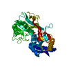
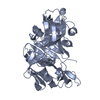
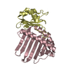
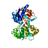
| Entry | Database: PDB / ID: 1h45 | ||||||
|---|---|---|---|---|---|---|---|
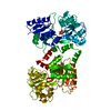
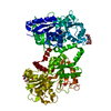
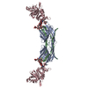
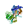
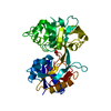
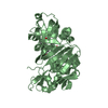
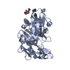
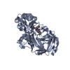
| Title | R210G N-TERMINAL LOBE HUMAN LACTOFERRIN | ||||||
 Components Components | LACTOFERRIN | ||||||
 Keywords Keywords | METAL TRANSPORT / IRON TRANSPORT / METAL BINDING | ||||||
| Function / homology |  Function and homology information Function and homology informationmembrane destabilizing activity / host-mediated suppression of viral proces / Mtb iron assimilation by chelation / phagocytic vesicle lumen / Metal sequestration by antimicrobial proteins / negative regulation of viral process / negative regulation of tumor necrosis factor (ligand) superfamily member 11 production / positive regulation of toll-like receptor 4 signaling pathway / negative regulation of single-species biofilm formation in or on host organism / positive regulation of bone mineralization involved in bone maturation ...membrane destabilizing activity / host-mediated suppression of viral proces / Mtb iron assimilation by chelation / phagocytic vesicle lumen / Metal sequestration by antimicrobial proteins / negative regulation of viral process / negative regulation of tumor necrosis factor (ligand) superfamily member 11 production / positive regulation of toll-like receptor 4 signaling pathway / negative regulation of single-species biofilm formation in or on host organism / positive regulation of bone mineralization involved in bone maturation / negative regulation of ATP-dependent activity / negative regulation of osteoclast development / specific granule / negative regulation of lipopolysaccharide-mediated signaling pathway / positive regulation of chondrocyte proliferation / antifungal humoral response / regulation of tumor necrosis factor production / bone morphogenesis / Antimicrobial peptides / positive regulation of osteoblast proliferation / negative regulation of viral genome replication / positive regulation of protein serine/threonine kinase activity / humoral immune response / Hydrolases; Acting on peptide bonds (peptidases); Serine endopeptidases / positive regulation of osteoblast differentiation / cysteine-type endopeptidase inhibitor activity / : / regulation of cytokine production / secretory granule / ossification / protein serine/threonine kinase activator activity / innate immune response in mucosa / iron ion transport / lipopolysaccharide binding / recycling endosome / specific granule lumen / tertiary granule lumen / antimicrobial humoral immune response mediated by antimicrobial peptide / heparin binding / antibacterial humoral response / killing of cells of another organism / defense response to Gram-negative bacterium / early endosome / positive regulation of canonical NF-kappaB signal transduction / iron ion binding / Amyloid fiber formation / serine-type endopeptidase activity / Neutrophil degranulation / negative regulation of apoptotic process / cell surface / protein-containing complex / proteolysis / : / DNA binding / extracellular exosome / extracellular region / nucleus / plasma membrane / cytoplasm Similarity search - Function | ||||||
| Biological species |  HOMO SAPIENS (human) HOMO SAPIENS (human) | ||||||
| Method |  X-RAY DIFFRACTION / MOLECULAR REPLACEMENT / Resolution: 1.95 Å X-RAY DIFFRACTION / MOLECULAR REPLACEMENT / Resolution: 1.95 Å | ||||||
 Authors Authors | Peterson, N.A. / Anderson, B.F. / Jameson, G.B. / Tweedie, J.W. / Baker, E.N. | ||||||
 Citation Citation | Journal: Biochemistry / Year: 2002 Title: "Dilysine Trigger" in Transferrins Probed by Mutagenesis of Lactoferrin: Crystal Structures of the R210G, R210E, and R210L Mutants of Human Lactoferrin Authors: Peterson, N.A. / Arcus, V. / Anderson, B.F. / Tweedie, J.W. / Jameson, G.B. / Baker, E.N. | ||||||
| History |
| ||||||
| Remark 700 | SHEET THE SHEET STRUCTURE OF THIS MOLECULE IS BIFURCATED. IN ORDER TO REPRESENT THIS FEATURE IN ... SHEET THE SHEET STRUCTURE OF THIS MOLECULE IS BIFURCATED. IN ORDER TO REPRESENT THIS FEATURE IN THE SHEET RECORDS BELOW, TWO SHEETS ARE DEFINED. |
- Structure visualization
| Structure viewer | Molecule: MolmilJmol/JSmol |
|---|
- Downloads & links
-Download
| PDBx/mmCIF format | 1h45.cif.gz | 78.3 KB | Display | PDBx/mmCIF format |
|---|---|---|---|---|
| PDB format | pdb1h45.ent.gz | 58 KB | Display | PDB format |
| PDBx/mmJSON format | 1h45.json.gz | Tree view | PDBx/mmJSON format | |
| Others |  Other downloads Other downloads |
-Validation report
| Arichive directory | https://data.pdbj.org/pub/pdb/validation_reports/h4/1h45ftp://data.pdbj.org/pub/pdb/validation_reports/h4/1h45 | HTTPS FTP |
|---|
-Related structure data
| Related structure data |  1h43C  1h44C  1lctS C: citing same article ( S: Starting model for refinement |
|---|---|
| Similar structure data |


-Links
 PDBj
PDBj


- Assembly
| Deposited unit | 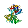
| ||||||||
|---|---|---|---|---|---|---|---|---|---|
| 1 |
| ||||||||
| Unit cell |
|
-Components
| #1: Protein | Mass: 37079.070 Da / Num. of mol.: 1 / Fragment: N-TERMINAL LOBE, RESIDUES 20-353 / Mutation: YES Source method: isolated from a genetically manipulated source Source: (gene. exp.) HOMO SAPIENS (human) / Cell line (production host): BHK / Production host:   CRICETULUS GRISEUS (Chinese hamster) / References: UniProt: P02788 CRICETULUS GRISEUS (Chinese hamster) / References: UniProt: P02788 |
|---|---|
| #2: Chemical | ChemComp-FE /   Mass: 55.845 Da / Num. of mol.: 1 / Source method: obtained synthetically / Formula: Fe Mass: 55.845 Da / Num. of mol.: 1 / Source method: obtained synthetically / Formula: Fe |
| #3: Chemical | ChemComp-CO3 /   Mass: 60.009 Da / Num. of mol.: 1 / Source method: obtained synthetically / Formula: CO3 Mass: 60.009 Da / Num. of mol.: 1 / Source method: obtained synthetically / Formula: CO3 |
| #4: Water | ChemComp-HOH /  Mass: 18.015 Da / Num. of mol.: 92 / Source method: isolated from a natural source / Formula: H2O Mass: 18.015 Da / Num. of mol.: 92 / Source method: isolated from a natural source / Formula: H2O |
| Compound details | IRON BINDING TRANSPORT PROTEINS THAT CAN BIND TWO FERRIC IRONS IN ASSOCIATION WITH THE BINDING OF ...IRON BINDING TRANSPORT PROTEINS THAT CAN BIND TWO FERRIC IRONS IN ASSOCIATIO |
| Has protein modification | Y |
| Sequence details | REGARDING THE SEQUENCE MISMATCH AT RESIDUE 28, THE SEQUENCE THAT WAS USED IN THIS WORK IS FROM THE ...REGARDING THE SEQUENCE MISMATCH AT RESIDUE 28, THE SEQUENCE THAT WAS USED IN THIS WORK IS FROM THE SEQUENCING |
-Experimental details
-Experiment
| Experiment | Method: X-RAY DIFFRACTION / Number of used crystals: 1 |
|---|
- Sample preparation
| Crystal | Density Matthews: 2.74 Å3/Da / Density % sol: 55.12 % | ||||||||||||||||||||||||||||
|---|---|---|---|---|---|---|---|---|---|---|---|---|---|---|---|---|---|---|---|---|---|---|---|---|---|---|---|---|---|
| Crystal grow | pH: 8 / Details: HEPES, NACL, pH 8.00 | ||||||||||||||||||||||||||||
| Crystal grow | *PLUS Temperature: 4 ℃ / pH: 8 / Method: batch method | ||||||||||||||||||||||||||||
| Components of the solutions | *PLUS
|
-Data collection
| Diffraction | Mean temperature: 113 K |
|---|---|
| Diffraction source | Source: ROTATING ANODE / Type: RIGAKU RU200 / Wavelength: 1.5418 |
| Detector | Type: RIGAKU IMAGE PLATE / Detector: IMAGE PLATE |
| Radiation | Monochromator: GRAPHITE / Protocol: SINGLE WAVELENGTH / Monochromatic (M) / Laue (L): M / Scattering type: x-ray |
| Radiation wavelength | Wavelength: 1.5418 Å / Relative weight: 1 |
| Reflection | Resolution: 1.95→30 Å / Num. obs: 27857 / % possible obs: 94.2 % / Redundancy: 2.8 % / Biso Wilson estimate: 17.8 Å2 / Rmerge(I) obs: 0.053 / Net I/σ(I): 13.6 |
| Reflection shell | Resolution: 1.95→2.02 Å / Rmerge(I) obs: 0.146 / Mean I/σ(I) obs: 4.2 / % possible all: 60.4 |
| Reflection | *PLUS Lowest resolution: 30 Å / Redundancy: 2.8 % |
| Reflection shell | *PLUS % possible obs: 60.4 % |
- Processing
| Software |
| ||||||||||||||||||||||||||||||||||||||||||||||||||||||||||||
|---|---|---|---|---|---|---|---|---|---|---|---|---|---|---|---|---|---|---|---|---|---|---|---|---|---|---|---|---|---|---|---|---|---|---|---|---|---|---|---|---|---|---|---|---|---|---|---|---|---|---|---|---|---|---|---|---|---|---|---|---|---|
| Refinement | Method to determine structure: MOLECULAR REPLACEMENT Starting model: PDB ENTRY 1LCT Resolution: 1.95→30 Å / Rfactor Rfree error: 0.007 / Isotropic thermal model: RESTRAINED / Cross valid method: THROUGHOUT / σ(F): 0
| ||||||||||||||||||||||||||||||||||||||||||||||||||||||||||||
| Solvent computation | Solvent model: FLAT MODEL / Bsol: 47.8612 Å2 / ksol: 0.313589 e/Å3 | ||||||||||||||||||||||||||||||||||||||||||||||||||||||||||||
| Displacement parameters | Biso mean: 33.2 Å2
| ||||||||||||||||||||||||||||||||||||||||||||||||||||||||||||
| Refine analyze |
| ||||||||||||||||||||||||||||||||||||||||||||||||||||||||||||
| Refinement step | Cycle: LAST / Resolution: 1.95→30 Å
| ||||||||||||||||||||||||||||||||||||||||||||||||||||||||||||
| Refine LS restraints |
| ||||||||||||||||||||||||||||||||||||||||||||||||||||||||||||
| LS refinement shell | Resolution: 1.95→2.07 Å / Rfactor Rfree error: 0.026 / Total num. of bins used: 6
| ||||||||||||||||||||||||||||||||||||||||||||||||||||||||||||
| Refinement | *PLUS Lowest resolution: 30 Å / Num. reflection obs: 26435 / Rfactor Rfree: 0.247 | ||||||||||||||||||||||||||||||||||||||||||||||||||||||||||||
| Solvent computation | *PLUS | ||||||||||||||||||||||||||||||||||||||||||||||||||||||||||||
| Displacement parameters | *PLUS | ||||||||||||||||||||||||||||||||||||||||||||||||||||||||||||
| Refine LS restraints | *PLUS
|
