 Movie
Movie Controller


+ Open data

- Basic information
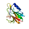
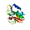
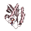
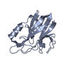
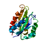
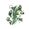
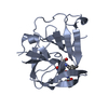
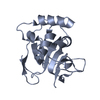
| Entry | Database: PDB / ID: 1gy2 | ||||||
|---|---|---|---|---|---|---|---|
| Title | Crystal structure of Met148Leu rusticyanin | ||||||
 Components Components | RUSTICYANIN | ||||||
 Keywords Keywords | ELECTRON TRANSPORT / S86D / M148L / RUSTICYANIN / MUTANT / METAL-BINDING / PERIPLASMIC | ||||||
| Function / homology |  Function and homology information Function and homology information | ||||||
| Biological species |  THIOBACILLUS FERROOXIDANS (bacteria) THIOBACILLUS FERROOXIDANS (bacteria) | ||||||
| Method |  X-RAY DIFFRACTION / SYNCHROTRON / MOLECULAR REPLACEMENT / Resolution: 1.82 Å X-RAY DIFFRACTION / SYNCHROTRON / MOLECULAR REPLACEMENT / Resolution: 1.82 Å | ||||||
 Authors Authors | Hough, M.A. / Kanbi, L.D. / Antonyuk, S. / Dodd, F. / Hasnain, S. | ||||||
 Citation Citation | Journal: J.Mol.Biol. / Year: 2002 Title: Crystal Structures of the met148Leu and Ser86Asp Mutants of Rusticyanin from Thiobacillus Ferrooxidans: Insights Into the Structural Relationship with the Cupredoxins and the Multi Copper Proteins. Authors: Kanbi, L.D. / Antonyuk, S. / Hough, M.A. / Hall, J.F. / Dodd, F. / Hasnain, S. | ||||||
| History |
|
- Structure visualization
| Structure viewer | Molecule: MolmilJmol/JSmol |
|---|
- Downloads & links
-Download
| PDBx/mmCIF format | 1gy2.cif.gz | 74.1 KB | Display | PDBx/mmCIF format |
|---|---|---|---|---|
| PDB format | pdb1gy2.ent.gz | 54.6 KB | Display | PDB format |
| PDBx/mmJSON format | 1gy2.json.gz | Tree view | PDBx/mmJSON format | |
| Others |  Other downloads Other downloads |
-Validation report
| Arichive directory | https://data.pdbj.org/pub/pdb/validation_reports/gy/1gy2ftp://data.pdbj.org/pub/pdb/validation_reports/gy/1gy2 | HTTPS FTP |
|---|
-Related structure data
| Related structure data |  1gy1C  1rcyS C: citing same article ( S: Starting model for refinement |
|---|---|
| Similar structure data |


-Links
 PDBj
PDBj- Assembly
| Deposited unit | 
| ||||||||
|---|---|---|---|---|---|---|---|---|---|
| 1 | 
| ||||||||
| 2 | 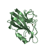
| ||||||||
| Unit cell |
|
-Components
| #1: Protein | Mass: 16550.898 Da / Num. of mol.: 2 / Mutation: YES Source method: isolated from a genetically manipulated source Source: (gene. exp.) THIOBACILLUS FERROOXIDANS (bacteria) / Production host: #2: Chemical |   Mass: 63.546 Da / Num. of mol.: 2 / Source method: obtained synthetically / Formula: Cu Mass: 63.546 Da / Num. of mol.: 2 / Source method: obtained synthetically / Formula: Cu#3: Chemical | ChemComp-GOL / |   Mass: 92.094 Da / Num. of mol.: 1 / Source method: obtained synthetically / Formula: C3H8O3 Mass: 92.094 Da / Num. of mol.: 1 / Source method: obtained synthetically / Formula: C3H8O3#4: Water | ChemComp-HOH / |  Mass: 18.015 Da / Num. of mol.: 136 / Source method: isolated from a natural source / Formula: H2O Mass: 18.015 Da / Num. of mol.: 136 / Source method: isolated from a natural source / Formula: H2OCompound details | CHAIN A, B ENGINEERED | |
|---|
-Experimental details
-Experiment
| Experiment | Method: X-RAY DIFFRACTION / Number of used crystals: 1 |
|---|
- Sample preparation
| Crystal | Density Matthews: 2.14 Å3/Da / Density % sol: 42.6 % | ||||||||||||||||||||||||||||||
|---|---|---|---|---|---|---|---|---|---|---|---|---|---|---|---|---|---|---|---|---|---|---|---|---|---|---|---|---|---|---|---|
| Crystal grow | pH: 5 / Details: 30% PEG 8000, 50 MM CITRIC ACID PH 5.0 | ||||||||||||||||||||||||||||||
| Crystal grow | *PLUS Temperature: 4 ℃ / pH: 3.8 / Method: vapor diffusion, hanging drop | ||||||||||||||||||||||||||||||
| Components of the solutions | *PLUS
|
-Data collection
| Diffraction | Mean temperature: 100 K |
|---|---|
| Diffraction source | Source: SYNCHROTRON / Site: SRS  / Beamline: PX9.5 / Wavelength: 1 / Beamline: PX9.5 / Wavelength: 1 |
| Detector | Type: ADSC CCD / Detector: CCD |
| Radiation | Protocol: SINGLE WAVELENGTH / Monochromatic (M) / Laue (L): M / Scattering type: x-ray |
| Radiation wavelength | Wavelength: 1 Å / Relative weight: 1 |
| Reflection | Resolution: 1.82→53.4 Å / Num. obs: 25330 / % possible obs: 92.2 % / Redundancy: 8.1 % / Rmerge(I) obs: 0.066 / Net I/σ(I): 15.1 |
| Reflection shell | Resolution: 1.82→1.84 Å / Redundancy: 3.1 % / Rmerge(I) obs: 0.345 / Mean I/σ(I) obs: 3 / % possible all: 94 |
| Reflection | *PLUS Num. measured all: 206007 |
| Reflection shell | *PLUS % possible obs: 94 % / Mean I/σ(I) obs: 2.96 |
- Processing
| Software |
| ||||||||||||||||||||
|---|---|---|---|---|---|---|---|---|---|---|---|---|---|---|---|---|---|---|---|---|---|
| Refinement | Method to determine structure: MOLECULAR REPLACEMENT Starting model: PDB ENTRY 1RCY Resolution: 1.82→53.4 Å / Cross valid method: THROUGHOUT / Details: NONE
| ||||||||||||||||||||
| Refinement step | Cycle: LAST / Resolution: 1.82→53.4 Å
| ||||||||||||||||||||
| Refinement | *PLUS Rfactor obs: 0.183 | ||||||||||||||||||||
| Solvent computation | *PLUS | ||||||||||||||||||||
| Displacement parameters | *PLUS | ||||||||||||||||||||
| Refine LS restraints | *PLUS
|
