 Movie
Movie Controller


+ Open data

- Basic information
| Entry | Database: PDB / ID: 1gwv | |||||||||
|---|---|---|---|---|---|---|---|---|---|---|
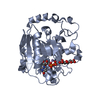
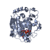
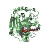
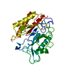
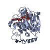
| Title | ALPHA-,1,3 GALACTOSYLTRANSFERASE - LACTOSE COMPLEX | |||||||||
 Components Components | N-ACETYLLACTOSAMINIDE ALPHA-1,3-GALACTOSYLTRANSFERASE | |||||||||
 Keywords Keywords | TRANSFERASE / GALACTOSYLTRANSFERASE / BLOOD GROUP SUGARS / LACTOSE / N-ACETYL LACTOSAMINE / GLYCOSYLTRANSFERASE | |||||||||
| Function / homology |  Function and homology information Function and homology informationN-acetyllactosaminide 3-alpha-galactosyltransferase / N-acetyllactosaminide 3-alpha-galactosyltransferase activity / Golgi cisterna / Golgi cisterna membrane / glycosyltransferase activity / carbohydrate metabolic process / vesicle / Golgi apparatus / metal ion binding Similarity search - Function | |||||||||
| Biological species |  | |||||||||
| Method |  X-RAY DIFFRACTION / SYNCHROTRON / MOLECULAR REPLACEMENT / Resolution: 2.5 Å X-RAY DIFFRACTION / SYNCHROTRON / MOLECULAR REPLACEMENT / Resolution: 2.5 Å | |||||||||
 Authors Authors | Boix, E. / Zhang, Y. / Swaminathan, G.J. / Brew, K. / Acharya, K.R. | |||||||||
 Citation Citation | Journal: J.Biol.Chem. / Year: 2002 Title: Structural Basis of Ordered Binding of Donor and Acceptor Substrates to the Retaining Glycosyltransferase, Alpha -1,3 Galactosyltransferase Authors: Boix, E. / Zhang, Y. / Swaminathan, G.J. / Brew, K. / Acharya, K.R. #1: Journal: J.Biol.Chem. / Year: 2001Title: Structure of Udp Complex of Udp-Galactose:Beta-Galactoside-Alpha -1,3-Galactosyltransferase at 1.53-A Resolution Reveals a Conformational Change in the Catalytically Important C Terminus Authors: Boix, E. / Swaminathan, G.J. / Zhang, Y. / Natesh, R. / Brew, K. / Acharya, K.R. | |||||||||
| History |
|
- Structure visualization
| Structure viewer | Molecule: MolmilJmol/JSmol |
|---|
- Downloads & links
-Download
| PDBx/mmCIF format | 1gwv.cif.gz | 138 KB | Display | PDBx/mmCIF format |
|---|---|---|---|---|
| PDB format | pdb1gwv.ent.gz | 105.3 KB | Display | PDB format |
| PDBx/mmJSON format | 1gwv.json.gz | Tree view | PDBx/mmJSON format | |
| Others |  Other downloads Other downloads |
-Validation report
| Arichive directory | https://data.pdbj.org/pub/pdb/validation_reports/gw/1gwvftp://data.pdbj.org/pub/pdb/validation_reports/gw/1gwv | HTTPS FTP |
|---|
-Related structure data
| Related structure data |  1gwwC  1gx0C  1gx4C  1k4vS C: citing same article ( S: Starting model for refinement |
|---|---|
| Similar structure data |


-Links
 PDBj
PDBj


- Assembly
| Deposited unit | 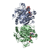
| ||||||||
|---|---|---|---|---|---|---|---|---|---|
| 1 | 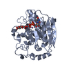
| ||||||||
| 2 | 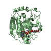
| ||||||||
| Unit cell |
|
-Components
| #1: Protein | Mass: 34090.164 Da / Num. of mol.: 2 / Fragment: CATALYTIC DOMAIN RESIDUES 80-368 Source method: isolated from a genetically manipulated source Source: (gene. exp.)  #2: Polysaccharide |   Source method: isolated from a genetically manipulated source Details: oligosaccharide / References: beta-lactose #3: Chemical |   Type: RNA linking / Mass: 404.161 Da / Num. of mol.: 2 / Source method: obtained synthetically / Formula: C9H14N2O12P2 / Comment: UDP*YM Type: RNA linking / Mass: 404.161 Da / Num. of mol.: 2 / Source method: obtained synthetically / Formula: C9H14N2O12P2 / Comment: UDP*YM#4: Chemical |   Mass: 54.938 Da / Num. of mol.: 2 / Source method: obtained synthetically / Formula: Mn Mass: 54.938 Da / Num. of mol.: 2 / Source method: obtained synthetically / Formula: Mn#5: Water | ChemComp-HOH / |  Mass: 18.015 Da / Num. of mol.: 180 / Source method: isolated from a natural source / Formula: H2O Mass: 18.015 Da / Num. of mol.: 180 / Source method: isolated from a natural source / Formula: H2O |
|---|
-Experimental details
-Experiment
| Experiment | Method: X-RAY DIFFRACTION / Number of used crystals: 1 |
|---|
- Sample preparation
| Crystal | Density Matthews: 2.6 Å3/Da / Density % sol: 57.59 % | ||||||||||||||||||||||||||||||||||||||||||||||||||||||||
|---|---|---|---|---|---|---|---|---|---|---|---|---|---|---|---|---|---|---|---|---|---|---|---|---|---|---|---|---|---|---|---|---|---|---|---|---|---|---|---|---|---|---|---|---|---|---|---|---|---|---|---|---|---|---|---|---|---|
| Crystal grow | pH: 8.5 / Details: PEG6000, TRIS/HCL, pH 8.50 | ||||||||||||||||||||||||||||||||||||||||||||||||||||||||
| Crystal grow | *PLUS Temperature: 16 ℃ / pH: 6 / Method: vapor diffusion, hanging drop | ||||||||||||||||||||||||||||||||||||||||||||||||||||||||
| Components of the solutions | *PLUS
|
-Data collection
| Diffraction | Mean temperature: 100 K |
|---|---|
| Diffraction source | Source: SYNCHROTRON / Site: SRS  / Beamline: PX14.1 / Wavelength: 1.488 / Beamline: PX14.1 / Wavelength: 1.488 |
| Detector | Type: ADSC CCD / Detector: CCD / Date: Apr 15, 2001 / Details: MIRRORS |
| Radiation | Protocol: SINGLE WAVELENGTH / Monochromatic (M) / Laue (L): M / Scattering type: x-ray |
| Radiation wavelength | Wavelength: 1.488 Å / Relative weight: 1 |
| Reflection | Resolution: 2.5→40 Å / Num. obs: 25766 / % possible obs: 95.3 % / Redundancy: 7.2 % / Biso Wilson estimate: 23.8 Å2 / Rmerge(I) obs: 0.126 / Net I/σ(I): 8.4 |
| Reflection shell | Resolution: 2.5→2.59 Å / Redundancy: 4.1 % / Rmerge(I) obs: 0.365 / Mean I/σ(I) obs: 3.01 / % possible all: 86.7 |
| Reflection | *PLUS Highest resolution: 2.5 Å / Lowest resolution: 39 Å / Num. obs: 27036 / Num. measured all: 82283 |
| Reflection shell | *PLUS Highest resolution: 2.5 Å / % possible obs: 86.7 % / Mean I/σ(I) obs: 2.9 |
- Processing
| Software |
| ||||||||||||||||||||||||||||||||||||||||||||||||||||||||||||||||||||||||||||||||
|---|---|---|---|---|---|---|---|---|---|---|---|---|---|---|---|---|---|---|---|---|---|---|---|---|---|---|---|---|---|---|---|---|---|---|---|---|---|---|---|---|---|---|---|---|---|---|---|---|---|---|---|---|---|---|---|---|---|---|---|---|---|---|---|---|---|---|---|---|---|---|---|---|---|---|---|---|---|---|---|---|---|
| Refinement | Method to determine structure: MOLECULAR REPLACEMENT Starting model: PDB ENTRY 1K4V Resolution: 2.5→38.98 Å / Rfactor Rfree error: 0.007 / Data cutoff high absF: 1087154.27 / Isotropic thermal model: RESTRAINED / Cross valid method: THROUGHOUT / σ(F): 0
| ||||||||||||||||||||||||||||||||||||||||||||||||||||||||||||||||||||||||||||||||
| Solvent computation | Solvent model: FLAT MODEL / Bsol: 22.8445 Å2 / ksol: 0.35378 e/Å3 | ||||||||||||||||||||||||||||||||||||||||||||||||||||||||||||||||||||||||||||||||
| Displacement parameters | Biso mean: 21.1 Å2
| ||||||||||||||||||||||||||||||||||||||||||||||||||||||||||||||||||||||||||||||||
| Refine analyze |
| ||||||||||||||||||||||||||||||||||||||||||||||||||||||||||||||||||||||||||||||||
| Refinement step | Cycle: LAST / Resolution: 2.5→38.98 Å
| ||||||||||||||||||||||||||||||||||||||||||||||||||||||||||||||||||||||||||||||||
| Refine LS restraints |
| ||||||||||||||||||||||||||||||||||||||||||||||||||||||||||||||||||||||||||||||||
| LS refinement shell | Resolution: 2.5→2.66 Å / Rfactor Rfree error: 0.024 / Total num. of bins used: 6
| ||||||||||||||||||||||||||||||||||||||||||||||||||||||||||||||||||||||||||||||||
| Xplor file |
| ||||||||||||||||||||||||||||||||||||||||||||||||||||||||||||||||||||||||||||||||
| Refinement | *PLUS Highest resolution: 2.5 Å / Lowest resolution: 39 Å / % reflection Rfree: 5 % | ||||||||||||||||||||||||||||||||||||||||||||||||||||||||||||||||||||||||||||||||
| Solvent computation | *PLUS | ||||||||||||||||||||||||||||||||||||||||||||||||||||||||||||||||||||||||||||||||
| Displacement parameters | *PLUS | ||||||||||||||||||||||||||||||||||||||||||||||||||||||||||||||||||||||||||||||||
| Refine LS restraints | *PLUS
| ||||||||||||||||||||||||||||||||||||||||||||||||||||||||||||||||||||||||||||||||
| LS refinement shell | *PLUS Highest resolution: 2.5 Å / Lowest resolution: 2.59 Å |
