 Movie
Movie Controller

[English] 日本語
 Yorodumi
- PDB-1vzt: ROLES OF INDIVIDUAL RESIDUES OF ALPHA-1,3 GALACTOSYLTRANSFERASES ... -
+ Open data

- Basic information
| Entry | Database: PDB / ID: 1vzt | |||||||||
|---|---|---|---|---|---|---|---|---|---|---|
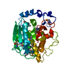
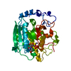
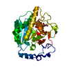
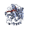
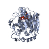
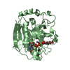
| Title | ROLES OF INDIVIDUAL RESIDUES OF ALPHA-1,3 GALACTOSYLTRANSFERASES IN SUBSTRATE BINDING AND CATALYSIS | |||||||||
 Components Components | N-ACETYLLACTOSAMINIDE ALPHA-1,3-GALACTOSYLTRANSFERASE | |||||||||
 Keywords Keywords | TRANSFERASE / ALPHA-1 / 3-GALACTOSYLTRANSFERASE-UDP COMPLEX / GLYCOSYLTRANSFERASE / GLYCOPROTEIN / TRANSMEMBRANE / NUCLEOTIDE-BINDING PROTEIN / XENOTRANSPLANTATION | |||||||||
| Function / homology |  Function and homology information Function and homology informationN-acetyllactosaminide 3-alpha-galactosyltransferase / N-acetyllactosaminide 3-alpha-galactosyltransferase activity / Golgi cisterna / Golgi cisterna membrane / glycosyltransferase activity / carbohydrate metabolic process / vesicle / Golgi apparatus / metal ion binding Similarity search - Function | |||||||||
| Biological species |  | |||||||||
| Method |  X-RAY DIFFRACTION / SYNCHROTRON / MOLECULAR REPLACEMENT / Resolution: 2 Å X-RAY DIFFRACTION / SYNCHROTRON / MOLECULAR REPLACEMENT / Resolution: 2 Å | |||||||||
 Authors Authors | Zhang, Y. / Swaminathan, G.J. / Deshpande, A. / Natesh, R. / Xie, X. / Acharya, K.R. / Brew, K. | |||||||||
 Citation Citation | Journal: Biochemistry / Year: 2003 Title: Roles of Individual Enzyme-Substrate Interactions by Alpha-1,3-Galactosyltransferase in Catalysis and Specificity. Authors: Zhang, Y. / Swaminathan, G.J. / Deshpande, A. / Boix, E. / Natesh, R. / Xie, Z. / Acharya, K.R. / Brew, K. #1: Journal: J.Biol.Chem. / Year: 2001Title: Udp-Galactose:Beta-Galactoside-Alpha-1,3-Galactosyl Transferase at1.53-A Resolution Reveals a Conformational Change in Thecatalytically Important C Terminus Authors: Boix, E. / Swaminathan, G.J. / Zhang, Y. / Natesh, R. / Brew, K. / Acharya, K.R. #2: Journal: Embo J. / Year: 2001Title: Bovine Alpha-1,3-Galactosyltransferases Catalytic Domain Structure and its Relationship with Abo Histo-Blood Group and Glycosphingolipid Glycosyltransferases Authors: Gastinel, L.N. / Bignon, C. / Misra, A.K. / Hindsgaul, O. / Shaper, J.H. / Joziasse, D.H. | |||||||||
| History |
| |||||||||
| Remark 700 | SHEET THE SHEET STRUCTURE OF THIS MOLECULE IS BIFURCATED. IN ORDER TO REPRESENT THIS FEATURE IN ... SHEET THE SHEET STRUCTURE OF THIS MOLECULE IS BIFURCATED. IN ORDER TO REPRESENT THIS FEATURE IN THE SHEET RECORDS BELOW, TWO SHEETS ARE DEFINED. |
- Structure visualization
| Structure viewer | Molecule: MolmilJmol/JSmol |
|---|
- Downloads & links
-Download
| PDBx/mmCIF format | 1vzt.cif.gz | 144.9 KB | Display | PDBx/mmCIF format |
|---|---|---|---|---|
| PDB format | pdb1vzt.ent.gz | 109.9 KB | Display | PDB format |
| PDBx/mmJSON format | 1vzt.json.gz | Tree view | PDBx/mmJSON format | |
| Others |  Other downloads Other downloads |
-Validation report
| Arichive directory | https://data.pdbj.org/pub/pdb/validation_reports/vz/1vztftp://data.pdbj.org/pub/pdb/validation_reports/vz/1vzt | HTTPS FTP |
|---|
-Related structure data
| Related structure data | 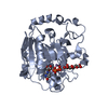 1o7oC  1o7qC  1k4vS S: Starting model for refinement C: citing same article ( |
|---|---|
| Similar structure data |


-Links
 PDBj
PDBj


- Assembly
| Deposited unit | 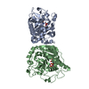
| ||||||||
|---|---|---|---|---|---|---|---|---|---|
| 1 | 
| ||||||||
| 2 | 
| ||||||||
| Unit cell |
|
-Components
| #1: Protein | Mass: 33961.004 Da / Num. of mol.: 2 / Fragment: CATALYTIC DOMAIN, RESIDUES 80-368 / Mutation: YES Source method: isolated from a genetically manipulated source Source: (gene. exp.)  #2: Chemical |   Mass: 54.938 Da / Num. of mol.: 2 / Source method: obtained synthetically / Formula: Mn Mass: 54.938 Da / Num. of mol.: 2 / Source method: obtained synthetically / Formula: Mn#3: Chemical |   Type: RNA linking / Mass: 404.161 Da / Num. of mol.: 2 / Source method: obtained synthetically / Formula: C9H14N2O12P2 / Comment: UDP*YM Type: RNA linking / Mass: 404.161 Da / Num. of mol.: 2 / Source method: obtained synthetically / Formula: C9H14N2O12P2 / Comment: UDP*YM#4: Chemical |   Mass: 122.143 Da / Num. of mol.: 2 / Source method: obtained synthetically / Formula: C4H12NO3 / Comment: pH buffer*YM Mass: 122.143 Da / Num. of mol.: 2 / Source method: obtained synthetically / Formula: C4H12NO3 / Comment: pH buffer*YM#5: Water | ChemComp-HOH / |  Mass: 18.015 Da / Num. of mol.: 442 / Source method: isolated from a natural source / Formula: H2O Mass: 18.015 Da / Num. of mol.: 442 / Source method: isolated from a natural source / Formula: H2OCompound details | TRANSFER OF GALACTOSE FROM UDP-GALACTOSE TO AN ACCEPTOR MOLECULE. UDP-GALACTOSE + BETA-D-GALACTOSYL- ...TRANSFER OF GALACTOSE FROM UDP-GALACTOSE TO AN ACCEPTOR MOLECULE. UDP-GALACTOSE + BETA-D-GALACTOSYL | |
|---|
-Experimental details
-Experiment
| Experiment | Method: X-RAY DIFFRACTION / Number of used crystals: 1 |
|---|
- Sample preparation
| Crystal | Density Matthews: 2.92 Å3/Da / Density % sol: 57.86 % |
|---|---|
| Crystal grow | pH: 8 / Details: PEG6000, TRIS/HCL, pH 8.00 |
-Data collection
| Diffraction | Mean temperature: 100 K |
|---|---|
| Diffraction source | Source: SYNCHROTRON / Site: SRS  / Beamline: PX14.1 / Wavelength: 1.488 / Beamline: PX14.1 / Wavelength: 1.488 |
| Detector | Type: ADSC CCD / Detector: CCD / Details: MIRRORS |
| Radiation | Protocol: SINGLE WAVELENGTH / Monochromatic (M) / Laue (L): M / Scattering type: x-ray |
| Radiation wavelength | Wavelength: 1.488 Å / Relative weight: 1 |
| Reflection | Resolution: 2→50 Å / Num. obs: 52584 / % possible obs: 92.2 % / Observed criterion σ(I): 0 / Redundancy: 3 % / Biso Wilson estimate: 22.93 Å2 / Net I/σ(I): 11.15 |
| Reflection shell | Resolution: 2→2.13 Å / % possible all: 84.5 |
- Processing
| Software |
| ||||||||||||||||||||||||||||||||||||||||||||||||||||||||||||
|---|---|---|---|---|---|---|---|---|---|---|---|---|---|---|---|---|---|---|---|---|---|---|---|---|---|---|---|---|---|---|---|---|---|---|---|---|---|---|---|---|---|---|---|---|---|---|---|---|---|---|---|---|---|---|---|---|---|---|---|---|---|
| Refinement | Method to determine structure: MOLECULAR REPLACEMENT Starting model: PDB ENTRY 1K4V Resolution: 2→50 Å / Rfactor Rfree error: 0.003 / Cross valid method: THROUGHOUT / σ(F): 0
| ||||||||||||||||||||||||||||||||||||||||||||||||||||||||||||
| Solvent computation | Bsol: 57.9859 Å2 / ksol: 0.400636 e/Å3 | ||||||||||||||||||||||||||||||||||||||||||||||||||||||||||||
| Displacement parameters | Biso mean: 57.91 Å2 | ||||||||||||||||||||||||||||||||||||||||||||||||||||||||||||
| Refine analyze |
| ||||||||||||||||||||||||||||||||||||||||||||||||||||||||||||
| Refinement step | Cycle: LAST / Resolution: 2→50 Å
| ||||||||||||||||||||||||||||||||||||||||||||||||||||||||||||
| Refine LS restraints |
| ||||||||||||||||||||||||||||||||||||||||||||||||||||||||||||
| LS refinement shell | Resolution: 2→2.13 Å / Rfactor Rfree error: 0.009 / Total num. of bins used: 6
| ||||||||||||||||||||||||||||||||||||||||||||||||||||||||||||
| Xplor file |
|
