 Movie
Movie Controller


+ Open data

- Basic information
| Entry | Database: PDB / ID: 1gvl | ||||||
|---|---|---|---|---|---|---|---|
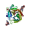
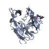
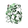
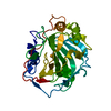
| Title | Human prokallikrein 6 (hK6)/ prozyme/ proprotease M/ proneurosin | ||||||
 Components Components | KALLIKREIN 6 | ||||||
 Keywords Keywords | HYDROLASE / ZYMOGEN / HUMAN KALLIKREIN | ||||||
| Function / homology |  Function and homology information Function and homology informationcornified envelope / tissue regeneration / positive regulation of G protein-coupled receptor signaling pathway / amyloid precursor protein metabolic process / hormone metabolic process / regulation of neuron projection development / protein autoprocessing / collagen catabolic process / regulation of cell differentiation / Hydrolases; Acting on peptide bonds (peptidases); Serine endopeptidases ...cornified envelope / tissue regeneration / positive regulation of G protein-coupled receptor signaling pathway / amyloid precursor protein metabolic process / hormone metabolic process / regulation of neuron projection development / protein autoprocessing / collagen catabolic process / regulation of cell differentiation / Hydrolases; Acting on peptide bonds (peptidases); Serine endopeptidases / intercellular bridge / myelination / secretory granule / central nervous system development / protein maturation / response to wounding / nuclear membrane / serine-type endopeptidase activity / nucleolus / mitochondrion / : / extracellular region / nucleoplasm / cytoplasm Similarity search - Function | ||||||
| Biological species |  HOMO SAPIENS (human) HOMO SAPIENS (human) | ||||||
| Method |  X-RAY DIFFRACTION / SYNCHROTRON / MOLECULAR REPLACEMENT / Resolution: 1.8 Å X-RAY DIFFRACTION / SYNCHROTRON / MOLECULAR REPLACEMENT / Resolution: 1.8 Å | ||||||
 Authors Authors | Gomis-Ruth, F.X. / Bayes, A. / Sotiropoulou, G. / Pampalakis, G. / Tsetsenis, T. / Villegas, V. / Aviles, F.X. / Coll, M. | ||||||
 Citation Citation | Journal: J.Biol.Chem. / Year: 2002 Title: The Structure of Human Prokallikrein 6 Reveals a Novel Activation Mechanism for the Kallikrein Family. Authors: Gomis-Ruth, F.X. / Bayes, A. / Sotiropoulou, G. / Pampalakis, G. / Tsetsenis, T. / Villegas, V. / Aviles, F.X. / Coll, M. | ||||||
| History |
| ||||||
| Remark 700 | SHEET DETERMINATION METHOD: DSSP THE SHEETS PRESENTED AS "AA" AND "AB" ON SHEET RECORDS BELOW ARE ... SHEET DETERMINATION METHOD: DSSP THE SHEETS PRESENTED AS "AA" AND "AB" ON SHEET RECORDS BELOW ARE ACTUALLY 7-STRANDED BARRELS. THESE ARE REPRESENTED BY 8-STRANDED SHEETS IN WHICH THE FIRST AND LAST STRANDS ARE IDENTICAL. |
- Structure visualization
| Structure viewer | Molecule: MolmilJmol/JSmol |
|---|
- Downloads & links
-Download
| PDBx/mmCIF format | 1gvl.cif.gz | 59.8 KB | Display | PDBx/mmCIF format |
|---|---|---|---|---|
| PDB format | pdb1gvl.ent.gz | 43 KB | Display | PDB format |
| PDBx/mmJSON format | 1gvl.json.gz | Tree view | PDBx/mmJSON format | |
| Others |  Other downloads Other downloads |
-Validation report
| Arichive directory | https://data.pdbj.org/pub/pdb/validation_reports/gv/1gvlftp://data.pdbj.org/pub/pdb/validation_reports/gv/1gvl | HTTPS FTP |
|---|
-Related structure data
| Related structure data | 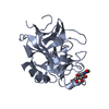 1npmS S: Starting model for refinement |
|---|---|
| Similar structure data |


-Links
 PDBj
PDBj- Assembly
| Deposited unit | 
| ||||||||
|---|---|---|---|---|---|---|---|---|---|
| 1 |
| ||||||||
| Unit cell |
|
-Components
| #1: Protein | Mass: 24517.838 Da / Num. of mol.: 1 / Mutation: YES Source method: isolated from a genetically manipulated source Source: (gene. exp.) HOMO SAPIENS (human) / Production host:  PICHIA PASTORIS (fungus) / Strain (production host): PPIC9 / References: UniProt: Q92876 PICHIA PASTORIS (fungus) / Strain (production host): PPIC9 / References: UniProt: Q92876 | ||
|---|---|---|---|
| #2: Water | ChemComp-HOH /  Mass: 18.015 Da / Num. of mol.: 158 / Source method: isolated from a natural source / Formula: H2O Mass: 18.015 Da / Num. of mol.: 158 / Source method: isolated from a natural source / Formula: H2O | ||
| Compound details | ENGINEERED| Has protein modification | Y | |
-Experimental details
-Experiment
| Experiment | Method: X-RAY DIFFRACTION / Number of used crystals: 1 |
|---|
- Sample preparation
| Crystal | Density Matthews: 2.2 Å3/Da / Density % sol: 43 % | |||||||||||||||||||||||||||||||||||||||||||||||||
|---|---|---|---|---|---|---|---|---|---|---|---|---|---|---|---|---|---|---|---|---|---|---|---|---|---|---|---|---|---|---|---|---|---|---|---|---|---|---|---|---|---|---|---|---|---|---|---|---|---|---|
| Crystal grow | *PLUS pH: 8.5 / Method: vapor diffusion, sitting drop | |||||||||||||||||||||||||||||||||||||||||||||||||
| Components of the solutions | *PLUS
|
-Data collection
| Diffraction | Mean temperature: 100 K |
|---|---|
| Diffraction source | Source: SYNCHROTRON / Site: ESRF  / Beamline: BM14 / Wavelength: 0.934 / Beamline: BM14 / Wavelength: 0.934 |
| Detector | Type: ADSC/MAR RESEARCH / Detector: CCD |
| Radiation | Protocol: SINGLE WAVELENGTH / Monochromatic (M) / Laue (L): M / Scattering type: x-ray |
| Radiation wavelength | Wavelength: 0.934 Å / Relative weight: 1 |
| Reflection | Resolution: 1.8→38.6 Å / Num. obs: 19445 / % possible obs: 94 % / Redundancy: 3.3 % / Biso Wilson estimate: 20 Å2 / Rmerge(I) obs: 0.095 / Net I/σ(I): 4.6 |
| Reflection shell | Resolution: 1.8→1.9 Å / Redundancy: 3 % / Rmerge(I) obs: 0.436 / Mean I/σ(I) obs: 1.7 / % possible all: 90.1 |
| Reflection | *PLUS Highest resolution: 1.8 Å / Num. measured all: 64722 |
| Reflection shell | *PLUS Highest resolution: 1.8 Å / Lowest resolution: 1.9 Å / % possible obs: 90.1 % |
- Processing
| Software |
| ||||||||||||||||||||||||||||||||||||||||||||||||||||||||||||
|---|---|---|---|---|---|---|---|---|---|---|---|---|---|---|---|---|---|---|---|---|---|---|---|---|---|---|---|---|---|---|---|---|---|---|---|---|---|---|---|---|---|---|---|---|---|---|---|---|---|---|---|---|---|---|---|---|---|---|---|---|---|
| Refinement | Method to determine structure: MOLECULAR REPLACEMENT Starting model: 1NPM Resolution: 1.8→33.2 Å / Isotropic thermal model: RESTRAINED / Cross valid method: THROUGHOUT / σ(F): 0 / Details: CHYMOTRYPSINOGEN A (CTGA) NUMBERING APPLIED.
| ||||||||||||||||||||||||||||||||||||||||||||||||||||||||||||
| Refinement step | Cycle: LAST / Resolution: 1.8→33.2 Å
| ||||||||||||||||||||||||||||||||||||||||||||||||||||||||||||
| Refine LS restraints |
| ||||||||||||||||||||||||||||||||||||||||||||||||||||||||||||
| Refinement | *PLUS % reflection Rfree: 7 % | ||||||||||||||||||||||||||||||||||||||||||||||||||||||||||||
| Solvent computation | *PLUS | ||||||||||||||||||||||||||||||||||||||||||||||||||||||||||||
| Displacement parameters | *PLUS |
