 Movie
Movie Controller

[English] 日本語
 Yorodumi
- PDB-3hzs: S. aureus monofunctional glycosyltransferase (MtgA)in complex wit... -
+ Open data

- Basic information
| Entry | Database: PDB / ID: 3hzs | ||||||
|---|---|---|---|---|---|---|---|
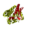
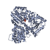
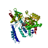
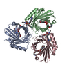
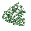
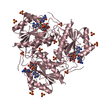
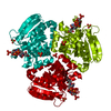
| Title | S. aureus monofunctional glycosyltransferase (MtgA)in complex with moenomycin | ||||||
 Components Components | Monofunctional glycosyltransferase | ||||||
 Keywords Keywords | TRANSFERASE / transglycosylase / peptidoglycan / monofunctional / moenomycin / Cell membrane / Cell shape / Cell wall biogenesis/degradation / Glycosyltransferase / Membrane / Peptidoglycan synthesis / Transmembrane | ||||||
| Function / homology |  Function and homology information Function and homology informationpeptidoglycan glycosyltransferase / peptidoglycan glycosyltransferase activity / peptidoglycan biosynthetic process / cell wall organization / regulation of cell shape / outer membrane-bounded periplasmic space / plasma membrane Similarity search - Function | ||||||
| Biological species |   Staphylococcus aureus subsp. aureus (bacteria) Staphylococcus aureus subsp. aureus (bacteria) | ||||||
| Method |  X-RAY DIFFRACTION / MOLECULAR REPLACEMENT / Resolution: 2.1 Å X-RAY DIFFRACTION / MOLECULAR REPLACEMENT / Resolution: 2.1 Å | ||||||
 Authors Authors | Heaslet, H. / Miller, A.A. / Shaw, B. / Mistry, A. | ||||||
 Citation Citation | Journal: J.Struct.Biol. / Year: 2009 Title: Characterization of the active site of S. aureus monofunctional glycosyltransferase (Mtg) by site-directed mutation and structural analysis of the protein complexed with moenomycin Authors: Heaslet, H. / Shaw, B. / Mistry, A. / Miller, A.A. | ||||||
| History |
|
- Structure visualization
| Structure viewer | Molecule: MolmilJmol/JSmol |
|---|
- Downloads & links
-Download
| PDBx/mmCIF format | 3hzs.cif.gz | 63.7 KB | Display | PDBx/mmCIF format |
|---|---|---|---|---|
| PDB format | pdb3hzs.ent.gz | 46.6 KB | Display | PDB format |
| PDBx/mmJSON format | 3hzs.json.gz | Tree view | PDBx/mmJSON format | |
| Others |  Other downloads Other downloads |
-Validation report
| Arichive directory | https://data.pdbj.org/pub/pdb/validation_reports/hz/3hzsftp://data.pdbj.org/pub/pdb/validation_reports/hz/3hzs | HTTPS FTP |
|---|
-Related structure data
| Related structure data | |
|---|---|
| Similar structure data |


-Links
 PDBj
PDBj- Assembly
| Deposited unit | 
| ||||||||
|---|---|---|---|---|---|---|---|---|---|
| 1 | 
| ||||||||
| Unit cell |
|
-Components
| #1: Protein | Mass: 24260.881 Da / Num. of mol.: 1 / Fragment: Staph. aureus monofunctional transglycosylase / Mutation: E100Q Source method: isolated from a genetically manipulated source Source: (gene. exp.) Staphylococcus aureus subsp. aureus (bacteria)Strain: MW2 / Gene: mgt, MW1814 / Plasmid: pPW2-SA0933(2)-N3 / Production host: References: UniProt: Q7A0I6, Transferases; Glycosyltransferases |
|---|---|
| #2: Chemical | ChemComp-M0E /   Mass: 1580.567 Da / Num. of mol.: 1 / Source method: obtained synthetically / Formula: C69H106N5O34P / Comment: antibiotic*YM Mass: 1580.567 Da / Num. of mol.: 1 / Source method: obtained synthetically / Formula: C69H106N5O34P / Comment: antibiotic*YM |
| #3: Chemical | ChemComp-PO4 /   Mass: 94.971 Da / Num. of mol.: 1 / Source method: obtained synthetically / Formula: PO4 Mass: 94.971 Da / Num. of mol.: 1 / Source method: obtained synthetically / Formula: PO4 |
| #4: Water | ChemComp-HOH /  Mass: 18.015 Da / Num. of mol.: 124 / Source method: isolated from a natural source / Formula: H2O Mass: 18.015 Da / Num. of mol.: 124 / Source method: isolated from a natural source / Formula: H2O |
-Experimental details
-Experiment
| Experiment | Method: X-RAY DIFFRACTION / Number of used crystals: 1 |
|---|
- Sample preparation
| Crystal | Density Matthews: 3.35 Å3/Da / Density % sol: 63.23 % |
|---|---|
| Crystal grow | Temperature: 295 K / Method: vapor diffusion, sitting drop / pH: 4.6 Details: Sa MtgA E100Q at 10 mg/ml was mixed with 1mM Moenomycin and 1mM MnCl2 and incubated on ice for ~3 hours. Precipitated material was removed by centrifugation at 16,000xg for 5 minutes. The ...Details: Sa MtgA E100Q at 10 mg/ml was mixed with 1mM Moenomycin and 1mM MnCl2 and incubated on ice for ~3 hours. Precipitated material was removed by centrifugation at 16,000xg for 5 minutes. The Mmoenomycin complex was crystallized by hanging drop vapor diffusion, mixing the protein 1:1 with a reservoir solution containing 0.1M Na Acetate pH 4.6, 0.2M NaCl, 30% MPD at 22oC. Hexagonal plate crystals formed in 1 week., VAPOR DIFFUSION, SITTING DROP, temperature 295K |
-Data collection
| Diffraction | Mean temperature: 100 K |
|---|---|
| Diffraction source | Source: ROTATING ANODE / Type: RIGAKU FR-E DW / Wavelength: 1.54 Å |
| Detector | Type: RIGAKU SATURN 944 / Detector: CCD / Date: Sep 12, 2007 / Details: Rigaku Varimax HF optics |
| Radiation | Protocol: SINGLE WAVELENGTH / Monochromatic (M) / Laue (L): M / Scattering type: x-ray |
| Radiation wavelength | Wavelength: 1.54 Å / Relative weight: 1 |
| Reflection | Resolution: 2.1→23.12 Å / Num. obs: 45531 / % possible obs: 99.9 % / Observed criterion σ(F): 2 / Observed criterion σ(I): 2 / Redundancy: 3.9 % / Rsym value: 0.173 / Net I/σ(I): 13.6 |
| Reflection shell | Resolution: 2.1→2.2 Å / Redundancy: 3.8 % / Mean I/σ(I) obs: 3.2 / Num. unique all: 4606 / Rsym value: 0.381 / % possible all: 100 |
- Processing
| Software |
| |||||||||||||||||||||||||||||||||||||||||||||||||||||||||||||||||
|---|---|---|---|---|---|---|---|---|---|---|---|---|---|---|---|---|---|---|---|---|---|---|---|---|---|---|---|---|---|---|---|---|---|---|---|---|---|---|---|---|---|---|---|---|---|---|---|---|---|---|---|---|---|---|---|---|---|---|---|---|---|---|---|---|---|---|
| Refinement | Method to determine structure: MOLECULAR REPLACEMENT Starting model: Unpublished apo structure of MTGA Resolution: 2.1→23.12 Å / Cor.coef. Fo:Fc: 0.957 / Cor.coef. Fo:Fc free: 0.926 / SU B: 4.49 / SU ML: 0.12 / Cross valid method: THROUGHOUT / σ(F): 2 / σ(I): 2 / ESU R: 0.178 / ESU R Free: 0.172 / Stereochemistry target values: MAXIMUM LIKELIHOOD / Details: HYDROGENS HAVE BEEN ADDED IN THE RIDING POSITIONS
| |||||||||||||||||||||||||||||||||||||||||||||||||||||||||||||||||
| Solvent computation | Ion probe radii: 0.8 Å / Shrinkage radii: 0.8 Å / VDW probe radii: 1.2 Å / Solvent model: MASK | |||||||||||||||||||||||||||||||||||||||||||||||||||||||||||||||||
| Displacement parameters | Biso mean: 36.033 Å2
| |||||||||||||||||||||||||||||||||||||||||||||||||||||||||||||||||
| Refinement step | Cycle: LAST / Resolution: 2.1→23.12 Å
| |||||||||||||||||||||||||||||||||||||||||||||||||||||||||||||||||
| Refine LS restraints |
| |||||||||||||||||||||||||||||||||||||||||||||||||||||||||||||||||
| LS refinement shell | Resolution: 2.1→2.155 Å / Total num. of bins used: 20
|
