 Movie
Movie Controller

[English] 日本語
 Yorodumi
- PDB-4fn8: Crystal structure of the Mtb enoyl CoA isomerase (Rv0632c)in comp... -
+ Open data

- Basic information
| Entry | Database: PDB / ID: 4fn8 | ||||||
|---|---|---|---|---|---|---|---|
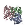
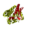
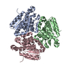
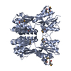
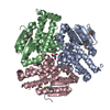
| Title | Crystal structure of the Mtb enoyl CoA isomerase (Rv0632c)in complex with acetoacetyl CoA | ||||||
 Components Components | Enoyl-CoA hydratase/isomerase family protein | ||||||
 Keywords Keywords | ISOMERASE / Structural Genomics / TB Structural Genomics Consortium / TBSGC / crotonase superfamily | ||||||
| Function / homology |  Function and homology information Function and homology informationenoyl-CoA hydratase / enoyl-CoA hydratase activity / fatty acid beta-oxidation / plasma membrane / cytosol Similarity search - Function | ||||||
| Biological species |   Mycobacterium tuberculosis (bacteria) Mycobacterium tuberculosis (bacteria) | ||||||
| Method |  X-RAY DIFFRACTION / MOLECULAR REPLACEMENT / molecular replacement / Resolution: 1.831 Å X-RAY DIFFRACTION / MOLECULAR REPLACEMENT / molecular replacement / Resolution: 1.831 Å | ||||||
 Authors Authors | Bruning, J.B. / Gao, N. / Hernandez, E.D. / Li, H. / Dang, N. / Hung, L.W. / Sacchettini, J.C. / TB Structural Genomics Consortium (TBSGC) | ||||||
 Citation Citation | Journal: To be Published Title: Crystal structure and mechanism of the prokaryotic enoyl CoA isomerase (ECI) Authors: Bruning, J.B. / Gao, N. / Hernandez, E.D. / Li, H. / Dang, N. / Hung, L.W. / Moran, S. / Sacchettini, J.C. | ||||||
| History |
|
- Structure visualization
| Structure viewer | Molecule: MolmilJmol/JSmol |
|---|
- Downloads & links
-Download
| PDBx/mmCIF format | 4fn8.cif.gz | 69.9 KB | Display | PDBx/mmCIF format |
|---|---|---|---|---|
| PDB format | pdb4fn8.ent.gz | 48.9 KB | Display | PDB format |
| PDBx/mmJSON format | 4fn8.json.gz | Tree view | PDBx/mmJSON format | |
| Others |  Other downloads Other downloads |
-Validation report
| Arichive directory | https://data.pdbj.org/pub/pdb/validation_reports/fn/4fn8ftp://data.pdbj.org/pub/pdb/validation_reports/fn/4fn8 | HTTPS FTP |
|---|
-Related structure data
| Related structure data |  4fn7S S: Starting model for refinement |
|---|---|
| Similar structure data | |
| Other databases |


-Links
 PDBj
PDBj
- Assembly
| Deposited unit | 
| ||||||||||||||||||||||||||||||||||||
|---|---|---|---|---|---|---|---|---|---|---|---|---|---|---|---|---|---|---|---|---|---|---|---|---|---|---|---|---|---|---|---|---|---|---|---|---|---|
| 1 | 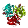
| ||||||||||||||||||||||||||||||||||||
| 2 | x 6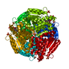
| ||||||||||||||||||||||||||||||||||||
| Unit cell |
| ||||||||||||||||||||||||||||||||||||
| Components on special symmetry positions |
|
-Components
| #1: Protein | Mass: 24466.951 Da / Num. of mol.: 1 Source method: isolated from a genetically manipulated source Source: (gene. exp.) Mycobacterium tuberculosis (bacteria) / Strain: H37Rv / Gene: echA3, MT0660, Rv0632c / Plasmid: pvp16 / Production host: | ||
|---|---|---|---|
| #2: Chemical | ChemComp-CAA /   Mass: 851.607 Da / Num. of mol.: 1 / Source method: obtained synthetically / Formula: C25H40N7O18P3S Mass: 851.607 Da / Num. of mol.: 1 / Source method: obtained synthetically / Formula: C25H40N7O18P3S | ||
| #3: Chemical |   Mass: 96.063 Da / Num. of mol.: 3 / Source method: obtained synthetically / Formula: SO4 Mass: 96.063 Da / Num. of mol.: 3 / Source method: obtained synthetically / Formula: SO4#4: Water | ChemComp-HOH / |  Mass: 18.015 Da / Num. of mol.: 367 / Source method: isolated from a natural source / Formula: H2O Mass: 18.015 Da / Num. of mol.: 367 / Source method: isolated from a natural source / Formula: H2O |
-Experimental details
-Experiment
| Experiment | Method: X-RAY DIFFRACTION / Number of used crystals: 1 |
|---|
- Sample preparation
| Crystal | Density Matthews: 2.3 Å3/Da / Density % sol: 46.54 % |
|---|---|
| Crystal grow | Temperature: 298 K / Method: vapor diffusion, hanging drop / pH: 6 Details: 1.6M ammonium sulfate, 0.1M MES 6.0, VAPOR DIFFUSION, HANGING DROP, temperature 298K |
-Data collection
| Diffraction | Mean temperature: 120 K | |||||||||||||||||||||||||||||||||||||||||||||||||||||||||||||||||||||||||||||||||||||||||||||||||||||||||||||||||||||||||||||||||||||||||||||||||||
|---|---|---|---|---|---|---|---|---|---|---|---|---|---|---|---|---|---|---|---|---|---|---|---|---|---|---|---|---|---|---|---|---|---|---|---|---|---|---|---|---|---|---|---|---|---|---|---|---|---|---|---|---|---|---|---|---|---|---|---|---|---|---|---|---|---|---|---|---|---|---|---|---|---|---|---|---|---|---|---|---|---|---|---|---|---|---|---|---|---|---|---|---|---|---|---|---|---|---|---|---|---|---|---|---|---|---|---|---|---|---|---|---|---|---|---|---|---|---|---|---|---|---|---|---|---|---|---|---|---|---|---|---|---|---|---|---|---|---|---|---|---|---|---|---|---|---|---|---|
| Diffraction source | Source: ROTATING ANODE / Type: RIGAKU RU200 / Wavelength: 1.54 Å | |||||||||||||||||||||||||||||||||||||||||||||||||||||||||||||||||||||||||||||||||||||||||||||||||||||||||||||||||||||||||||||||||||||||||||||||||||
| Detector | Type: RIGAKU RAXIS IV++ / Detector: IMAGE PLATE / Date: Jul 30, 2009 / Details: osmic mirrors | |||||||||||||||||||||||||||||||||||||||||||||||||||||||||||||||||||||||||||||||||||||||||||||||||||||||||||||||||||||||||||||||||||||||||||||||||||
| Radiation | Monochromator: osmic mirrors / Protocol: SINGLE WAVELENGTH / Monochromatic (M) / Laue (L): M / Scattering type: x-ray | |||||||||||||||||||||||||||||||||||||||||||||||||||||||||||||||||||||||||||||||||||||||||||||||||||||||||||||||||||||||||||||||||||||||||||||||||||
| Radiation wavelength | Wavelength: 1.54 Å / Relative weight: 1 | |||||||||||||||||||||||||||||||||||||||||||||||||||||||||||||||||||||||||||||||||||||||||||||||||||||||||||||||||||||||||||||||||||||||||||||||||||
| Reflection | Redundancy: 7.2 % / Av σ(I) over netI: 42.59 / Number: 151417 / Rmerge(I) obs: 0.06 / Χ2: 2.03 / D res high: 1.83 Å / D res low: 50 Å / Num. obs: 21089 / % possible obs: 99.9 | |||||||||||||||||||||||||||||||||||||||||||||||||||||||||||||||||||||||||||||||||||||||||||||||||||||||||||||||||||||||||||||||||||||||||||||||||||
| Diffraction reflection shell |
| |||||||||||||||||||||||||||||||||||||||||||||||||||||||||||||||||||||||||||||||||||||||||||||||||||||||||||||||||||||||||||||||||||||||||||||||||||
| Reflection | Resolution: 1.83→50 Å / Num. all: 21089 / Num. obs: 21089 / % possible obs: 99.9 % / Observed criterion σ(F): 0 / Observed criterion σ(I): 0 / Redundancy: 7.2 % / Biso Wilson estimate: 17.87 Å2 / Rmerge(I) obs: 0.06 / Rsym value: 0.06 / Χ2: 2.031 / Net I/σ(I): 17.4 | |||||||||||||||||||||||||||||||||||||||||||||||||||||||||||||||||||||||||||||||||||||||||||||||||||||||||||||||||||||||||||||||||||||||||||||||||||
| Reflection shell |
|
-Phasing
| Phasing | Method: molecular replacement |
|---|
- Processing
| Software |
| ||||||||||||||||||||||||||||||||||||||||||||||||||||||
|---|---|---|---|---|---|---|---|---|---|---|---|---|---|---|---|---|---|---|---|---|---|---|---|---|---|---|---|---|---|---|---|---|---|---|---|---|---|---|---|---|---|---|---|---|---|---|---|---|---|---|---|---|---|---|---|
| Refinement | Method to determine structure: MOLECULAR REPLACEMENT Starting model: PDB ENTRY 4FN7 subunit A Resolution: 1.831→25.35 Å / Occupancy max: 1 / Occupancy min: 0.12 / FOM work R set: 0.9014 / SU ML: 0.18 / Isotropic thermal model: isotropic / Cross valid method: THROUGHOUT / σ(F): 0 / σ(I): 0 / Phase error: 15.39 / Stereochemistry target values: ML
| ||||||||||||||||||||||||||||||||||||||||||||||||||||||
| Solvent computation | Shrinkage radii: 0.9 Å / VDW probe radii: 1.11 Å / Solvent model: FLAT BULK SOLVENT MODEL / Bsol: 74.986 Å2 / ksol: 0.365 e/Å3 | ||||||||||||||||||||||||||||||||||||||||||||||||||||||
| Displacement parameters | Biso max: 71.6 Å2 / Biso mean: 18.8567 Å2 / Biso min: 5.24 Å2
| ||||||||||||||||||||||||||||||||||||||||||||||||||||||
| Refinement step | Cycle: LAST / Resolution: 1.831→25.35 Å
| ||||||||||||||||||||||||||||||||||||||||||||||||||||||
| Refine LS restraints |
| ||||||||||||||||||||||||||||||||||||||||||||||||||||||
| LS refinement shell | Refine-ID: X-RAY DIFFRACTION / Total num. of bins used: 8 / % reflection obs: 100 %
|
