- PDB-1gvc: 18kDa N-II domain fragment of duck ovotransferrin + NTA -
+
Open data
ID or keywords:
Loading...
-
Basic information
Entry
Database: PDB / ID: 1gvc
Title
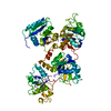
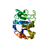
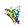
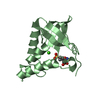
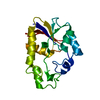
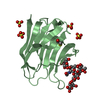
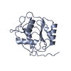
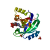
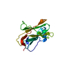
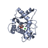
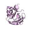
18kDa N-II domain fragment of duck ovotransferrin + NTA
Components
OVOTRANSFERRIN
Keywords
IRON TRANSPORT / GLYCOPROTEIN / METAL-BINDING
Function / homology
Function and homology information
iron ion transport / recycling endosome / antibacterial humoral response / early endosome / : / metal ion binding / plasma membrane Similarity search - Function
SHEET THE SHEET STRUCTURE OF THIS MOLECULE IS BIFURCATED. IN ORDER TO REPRESENT THIS FEATURE IN ... SHEET THE SHEET STRUCTURE OF THIS MOLECULE IS BIFURCATED. IN ORDER TO REPRESENT THIS FEATURE IN THE SHEET RECORDS BELOW, TWO SHEETS ARE DEFINED.
Mass: 17216.471 Da / Num. of mol.: 1 / Fragment: N-II FRAGMENT, RESIDUES 91-247 / Source method: isolated from a natural source / Details: COMPLEX OF DUCK OVOTRANSFERRIN FRAGMENT AND NTA / Source: (natural) ANAS PLATYRHYNCHOS (mallard) / References: UniProt: P56410
Mass: 18.015 Da / Num. of mol.: 96 / Source method: isolated from a natural source / Formula: H2O
Has protein modification
Y
Sequence details
SEQUENCED BY DR. R.W.EVANS (GUY'S HOSPITAL) LONDON, U.K. THESE AMINO-ACID RESIDUES DIFFER FROM ...SEQUENCED BY DR. R.W.EVANS (GUY'S HOSPITAL) LONDON, U.K. THESE AMINO-ACID RESIDUES DIFFER FROM THOSE GIVEN BY SWISS-PROT.
-
Experimental details
-
Experiment
Experiment
Method: X-RAY DIFFRACTION / Number of used crystals: 1
-
Sample preparation
Crystal
Density Matthews: 2.24 Å3/Da / Density % sol: 45 %
Crystal grow
Temperature: 277 K / Method: vapor diffusion, sitting drop / pH: 7.8 / Details: SITTING DROP AT PH7.8 AND 277K, pH 7.80
Crystal grow
*PLUS
Temperature: 277 K / Method: vapor diffusion, sitting drop
Type: RIGAKU RAXIS II / Detector: IMAGE PLATE / Date: Jan 15, 1993 / Details: PT COATED FUSED QUARTZ
Radiation
Monochromator: TRIANGULAR SI(111) / Protocol: SINGLE WAVELENGTH / Monochromatic (M) / Laue (L): M / Scattering type: x-ray
Radiation wavelength
Wavelength: 0.88 Å / Relative weight: 1
Reflection
Resolution: 1.9→26.92 Å / Num. obs: 12081 / % possible obs: 98 % / Redundancy: 4.3 % / Rmerge(I) obs: 0.041 / Net I/σ(I): 8.5
Reflection shell
Resolution: 1.9→1.97 Å / Redundancy: 2.1 % / Rmerge(I) obs: 0.088 / Mean I/σ(I) obs: 7.6 / % possible all: 97.3
Reflection
*PLUS
Num. measured all: 52160
Reflection shell
*PLUS
% possible obs: 97.3 %
-
Processing
Software
Name
Version
Classification
REFMAC
5
refinement
MOSFLM
datareduction
Agrovata
datascaling
Refinement
Method to determine structure: OTHER / Resolution: 1.9→26.92 Å / SU B: 4.873 / SU ML: 0.145 / Cross valid method: THROUGHOUT / ESU R Free: 0.139 / Details: RESIDUES E144 - S148 INCLUSIVE ARE POORLY DEFINED
Rfactor
Num. reflection
% reflection
Selection details
Rfree
0.211
573
4.8 %
RANDOM
Rwork
0.185
-
-
-
obs
0.186
11481
98.05 %
-
Refinement step
Cycle: LAST / Resolution: 1.9→26.92 Å
Protein
Nucleic acid
Ligand
Solvent
Total
Num. atoms
1204
0
18
96
1318
Software
*PLUS
Name: REFMAC / Classification: refinement
Refine LS restraints
*PLUS
Refine-ID
Type
Dev ideal
X-RAY DIFFRACTION
p_bond_d
0.013
X-RAY DIFFRACTION
p_angle_d
X-RAY DIFFRACTION
p_angle_deg
1.68
X-RAY DIFFRACTION
p_plane_restr
0.006
+
About Yorodumi
-
News
-
Feb 9, 2022. New format data for meta-information of EMDB entries
New format data for meta-information of EMDB entries
Version 3 of the EMDB header file is now the official format.
The previous official version 1.9 will be removed from the archive.
In the structure databanks used in Yorodumi, some data are registered as the other names, "COVID-19 virus" and "2019-nCoV". Here are the details of the virus and the list of structure data.
Jan 31, 2019. EMDB accession codes are about to change! (news from PDBe EMDB page)
EMDB accession codes are about to change! (news from PDBe EMDB page)
The allocation of 4 digits for EMDB accession codes will soon come to an end. Whilst these codes will remain in use, new EMDB accession codes will include an additional digit and will expand incrementally as the available range of codes is exhausted. The current 4-digit format prefixed with “EMD-” (i.e. EMD-XXXX) will advance to a 5-digit format (i.e. EMD-XXXXX), and so on. It is currently estimated that the 4-digit codes will be depleted around Spring 2019, at which point the 5-digit format will come into force.
The EM Navigator/Yorodumi systems omit the EMD- prefix.
Related info.:Q: What is EMD? / ID/Accession-code notation in Yorodumi/EM Navigator
Yorodumi is a browser for structure data from EMDB, PDB, SASBDB, etc.
This page is also the successor to EM Navigator detail page, and also detail information page/front-end page for Omokage search.
The word "yorodu" (or yorozu) is an old Japanese word meaning "ten thousand". "mi" (miru) is to see.
Related info.:EMDB / PDB / SASBDB / Comparison of 3 databanks / Yorodumi Search / Aug 31, 2016. New EM Navigator & Yorodumi / Yorodumi Papers / Jmol/JSmol / Function and homology information / Changes in new EM Navigator and Yorodumi
 Movie
Movie Controller


 Open data
 Basic information
 Components
 Keywords
 Function and homology information
 X-RAY DIFFRACTION /
  Authors
 Citation
 Structure visualization
 Downloads & links
 Other downloads


 PDBj
PDBj


 Assembly
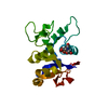

 Mass: 60.009 Da / Num. of mol.: 1 / Source method: obtained synthetically / Formula: CO3
 Mass: 191.139 Da / Num. of mol.: 1 / Source method: obtained synthetically / Formula: C6H9NO6
 Mass: 55.845 Da / Num. of mol.: 1 / Source method: obtained synthetically / Formula: Fe
 Mass: 18.015 Da / Num. of mol.: 96 / Source method: isolated from a natural source / Formula: H2O
 Sample preparation
 / Beamline: PX9.6 / Wavelength: 0.88
  Processing