 Movie
Movie Controller

[English] 日本語
 Yorodumi
- PDB-6xbt: Streptomyces coelicolor methylmalonyl-CoA epimerase (Q60A) in com... -
+ Open data

- Basic information
| Entry | Database: PDB / ID: 6xbt | ||||||
|---|---|---|---|---|---|---|---|
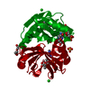
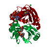
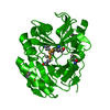
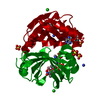
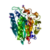
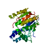
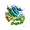
| Title | Streptomyces coelicolor methylmalonyl-CoA epimerase (Q60A) in complex with 2-nitronate-propionyl-CoA | ||||||
 Components Components | Methylmalonyl-CoA epimerase | ||||||
 Keywords Keywords | ISOMERASE/INHIBITOR / Methylmalonyl-CoA / epimerase / enol/enolate / enol/enolate analog / ISOMERASE / ISOMERASE-INHIBITOR complex | ||||||
| Function / homology |  Function and homology information Function and homology informationmethylmalonyl-CoA epimerase activity / L-methylmalonyl-CoA metabolic process / metal ion binding Similarity search - Function | ||||||
| Biological species |  Streptomyces coelicolor (bacteria) Streptomyces coelicolor (bacteria) | ||||||
| Method |  X-RAY DIFFRACTION / SYNCHROTRON / MOLECULAR REPLACEMENT / molecular replacement / Resolution: 1.49 Å X-RAY DIFFRACTION / SYNCHROTRON / MOLECULAR REPLACEMENT / molecular replacement / Resolution: 1.49 Å | ||||||
 Authors Authors | Stunkard, L.M. / Boram, T.J. / Benjamin, A.B. / Bower, J.B. / Lohman, J.R. | ||||||
 Citation Citation | Journal: To Be Published Title: Streptomyces coelicolor methylmalonyl-CoA epimerase (Q60A) in complex with 2-nitronate-propionyl-CoA Authors: Stunkard, L.M. / Boram, T.J. / Benjamin, A.B. / Bower, J.B. / Lohman, J.R. | ||||||
| History |
|
- Structure visualization
| Structure viewer | Molecule: MolmilJmol/JSmol |
|---|
- Downloads & links
-Download
| PDBx/mmCIF format | 6xbt.cif.gz | 56.7 KB | Display | PDBx/mmCIF format |
|---|---|---|---|---|
| PDB format | pdb6xbt.ent.gz | 36.9 KB | Display | PDB format |
| PDBx/mmJSON format | 6xbt.json.gz | Tree view | PDBx/mmJSON format | |
| Others |  Other downloads Other downloads |
-Validation report
| Arichive directory | https://data.pdbj.org/pub/pdb/validation_reports/xb/6xbtftp://data.pdbj.org/pub/pdb/validation_reports/xb/6xbt | HTTPS FTP |
|---|
-Related structure data
| Related structure data |  1jc5S S: Starting model for refinement |
|---|---|
| Similar structure data |

-Links
 PDBj
PDBj


- Assembly
| Deposited unit | 
| ||||||||
|---|---|---|---|---|---|---|---|---|---|
| 1 | 
| ||||||||
| Unit cell |
| ||||||||
| Components on special symmetry positions |
|
-Components
-Protein , 1 types, 1 molecules A
| #1: Protein | Mass: 16002.716 Da / Num. of mol.: 1 / Mutation: M1S, Q60A Source method: isolated from a genetically manipulated source Source: (gene. exp.) Streptomyces coelicolor (bacteria) / Strain: ATCC BAA-471 / A3(2) / M145 / Gene: SCO5398 / Production host: |
|---|
-Non-polymers , 5 types, 223 molecules 


| #2: Chemical | ChemComp-CO /  Mass: 58.933 Da / Num. of mol.: 1 / Source method: obtained synthetically / Formula: Co Mass: 58.933 Da / Num. of mol.: 1 / Source method: obtained synthetically / Formula: Co | ||||||
|---|---|---|---|---|---|---|---|
| #3: Chemical |  Mass: 96.063 Da / Num. of mol.: 2 / Source method: obtained synthetically / Formula: SO4 Mass: 96.063 Da / Num. of mol.: 2 / Source method: obtained synthetically / Formula: SO4#4: Chemical | ChemComp-KFV / [ |  Mass: 867.587 Da / Num. of mol.: 1 / Source method: obtained synthetically / Formula: C24H38N8O19P3S / Feature type: SUBJECT OF INVESTIGATION Mass: 867.587 Da / Num. of mol.: 1 / Source method: obtained synthetically / Formula: C24H38N8O19P3S / Feature type: SUBJECT OF INVESTIGATION#5: Chemical | ChemComp-COA / |  Mass: 767.534 Da / Num. of mol.: 1 / Source method: obtained synthetically / Formula: C21H36N7O16P3S Mass: 767.534 Da / Num. of mol.: 1 / Source method: obtained synthetically / Formula: C21H36N7O16P3S#6: Water | ChemComp-HOH / | Mass: 18.015 Da / Num. of mol.: 218 / Source method: isolated from a natural source / Formula: H2O |
-Details
| Has ligand of interest | Y |
|---|
-Experimental details
-Experiment
| Experiment | Method: X-RAY DIFFRACTION / Number of used crystals: 1 |
|---|
- Sample preparation
| Crystal | Density Matthews: 3.73 Å3/Da / Density % sol: 67.04 % |
|---|---|
| Crystal grow | Temperature: 298 K / Method: vapor diffusion, hanging drop / pH: 7 Details: 150 mM sodium chloride, 100 mM Bis-Tris:HCl pH 7.0, 2.3 M ammonium sulfate, and 5% PEG400 Temp details: room temperature |
-Data collection
| Diffraction | Mean temperature: 80 K / Ambient temp details: liquid nitrogen / Serial crystal experiment: N |
|---|---|
| Diffraction source | Source: SYNCHROTRON / Site: APS  / Beamline: 21-ID-G / Wavelength: 0.97856 Å / Beamline: 21-ID-G / Wavelength: 0.97856 Å |
| Detector | Type: MAR scanner 300 mm plate / Detector: IMAGE PLATE / Date: Apr 5, 2019 / Details: MD2 microdifractometer |
| Radiation | Monochromator: C(111) / Protocol: SINGLE WAVELENGTH / Monochromatic (M) / Laue (L): M / Scattering type: x-ray |
| Radiation wavelength | Wavelength: 0.97856 Å / Relative weight: 1 |
| Reflection | Resolution: 1.49→30 Å / Num. obs: 40894 / % possible obs: 100 % / Redundancy: 14 % / Biso Wilson estimate: 27.36 Å2 / CC1/2: 0.985 / CC star: 0.996 / Rmerge(I) obs: 0.1 / Rpim(I) all: 0.028 / Rrim(I) all: 0.104 / Rsym value: 0.091 / Χ2: 1.046 / Net I/σ(I): 446.2 |
| Reflection shell | Resolution: 1.49→1.55 Å / Redundancy: 13.9 % / Rmerge(I) obs: 0.846 / Mean I/σ(I) obs: 8.6 / Num. unique obs: 3986 / CC1/2: 0.905 / CC star: 0.975 / Rpim(I) all: 0.234 / Rrim(I) all: 0.878 / Rsym value: 0.708 / Χ2: 0.936 / % possible all: 100 |
-Phasing
| Phasing | Method: molecular replacement | |||||||||
|---|---|---|---|---|---|---|---|---|---|---|
| Phasing MR | Model details: Phaser MODE: MR_AUTO
|
- Processing
| Software |
| ||||||||||||||||||||||||||||||||||||||||||||||||||||||||||||
|---|---|---|---|---|---|---|---|---|---|---|---|---|---|---|---|---|---|---|---|---|---|---|---|---|---|---|---|---|---|---|---|---|---|---|---|---|---|---|---|---|---|---|---|---|---|---|---|---|---|---|---|---|---|---|---|---|---|---|---|---|---|
| Refinement | Method to determine structure: MOLECULAR REPLACEMENT Starting model: 1JC5 Resolution: 1.49→29.5 Å / Cor.coef. Fo:Fc: 0.972 / Cor.coef. Fo:Fc free: 0.972 / SU B: 1.011 / SU ML: 0.037 / SU R Cruickshank DPI: 0.0547 / Cross valid method: THROUGHOUT / σ(F): 0 / ESU R: 0.055 / ESU R Free: 0.055 / Stereochemistry target values: MAXIMUM LIKELIHOOD Details: HYDROGENS HAVE BEEN ADDED IN THE RIDING POSITIONS U VALUES : REFINED INDIVIDUALLY
| ||||||||||||||||||||||||||||||||||||||||||||||||||||||||||||
| Solvent computation | Ion probe radii: 0.8 Å / Shrinkage radii: 0.8 Å / VDW probe radii: 1.2 Å / Solvent model: MASK | ||||||||||||||||||||||||||||||||||||||||||||||||||||||||||||
| Displacement parameters | Biso max: 106.08 Å2 / Biso mean: 27.36 Å2 / Biso min: 13.37 Å2
| ||||||||||||||||||||||||||||||||||||||||||||||||||||||||||||
| Refinement step | Cycle: final / Resolution: 1.49→29.5 Å
| ||||||||||||||||||||||||||||||||||||||||||||||||||||||||||||
| Refine LS restraints |
| ||||||||||||||||||||||||||||||||||||||||||||||||||||||||||||
| LS refinement shell | Resolution: 1.493→1.532 Å / Rfactor Rfree error: 0 / Total num. of bins used: 20
|
