 Movie
Movie Controller


+ Open data

- Basic information
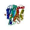
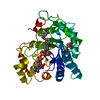
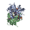
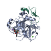
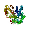
| Entry | Database: PDB / ID: 1jc5 | ||||||
|---|---|---|---|---|---|---|---|
| Title | Crystal Structure of Native Methylmalonyl-CoA Epimerase | ||||||
 Components Components | Methylmalonyl-CoA Epimerase | ||||||
 Keywords Keywords | ISOMERASE / epimerisation / methylmalonyl-CoA / vicinal oxygen chelate superfamily / metal-assisted catalysis | ||||||
| Function / homology |  Function and homology information Function and homology informationmethylmalonyl-CoA epimerase / methylmalonyl-CoA epimerase activity / L-methylmalonyl-CoA metabolic process / metal ion binding Similarity search - Function | ||||||
| Biological species |  Propionibacterium freudenreichii subsp. shermanii (bacteria) Propionibacterium freudenreichii subsp. shermanii (bacteria) | ||||||
| Method |  X-RAY DIFFRACTION / SYNCHROTRON / MOLECULAR REPLACEMENT / Resolution: 2.2 Å X-RAY DIFFRACTION / SYNCHROTRON / MOLECULAR REPLACEMENT / Resolution: 2.2 Å | ||||||
 Authors Authors | Mc Carthy, A.A. / Baker, H.M. / Shewry, S.C. / Patchett, M.L. / Baker, E.N. | ||||||
 Citation Citation | Journal: Structure / Year: 2001 Title: Crystal structure of methylmalonyl-coenzyme A epimerase from P. shermanii: a novel enzymatic function on an ancient metal binding scaffold. Authors: McCarthy, A.A. / Baker, H.M. / Shewry, S.C. / Patchett, M.L. / Baker, E.N. #1: Journal: Acta Crystallogr.,Sect.D / Year: 2001Title: Expression, Crystallization and Preliminary Characterization of Methylmalonyl Coenzyme A Epimerase from Propionibacterium shermanii Authors: Mc Carthy, A.A. / Baker, H.M. / Shewry, S.C. / Kagawa, T.F. / Saafi, E. / Patchett, M.L. / Baker, E.N. | ||||||
| History |
|
- Structure visualization
| Structure viewer | Molecule: MolmilJmol/JSmol |
|---|
- Downloads & links
-Download
| PDBx/mmCIF format | 1jc5.cif.gz | 179.9 KB | Display | PDBx/mmCIF format |
|---|---|---|---|---|
| PDB format | pdb1jc5.ent.gz | 145.6 KB | Display | PDB format |
| PDBx/mmJSON format | 1jc5.json.gz | Tree view | PDBx/mmJSON format | |
| Others |  Other downloads Other downloads |
-Validation report
| Arichive directory | https://data.pdbj.org/pub/pdb/validation_reports/jc/1jc5ftp://data.pdbj.org/pub/pdb/validation_reports/jc/1jc5 | HTTPS FTP |
|---|
-Related structure data


-Links
 PDBj
PDBj- Assembly
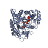
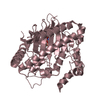
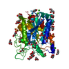
| Deposited unit | 
| ||||||||
|---|---|---|---|---|---|---|---|---|---|
| 1 | 
| ||||||||
| 2 | 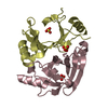
| ||||||||
| 3 | 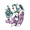
| ||||||||
| Unit cell |
| ||||||||
| Details | The biological assembly is a homodimer |
-Components
| #1: Protein | Mass: 16738.908 Da / Num. of mol.: 6 Source method: isolated from a genetically manipulated source Source: (gene. exp.) Propionibacterium freudenreichii subsp. shermanii (bacteria)Species: Propionibacterium freudenreichii / Strain: subsp. shermanii / Plasmid: pTEEX / Species (production host): Escherichia coli / Production host: #2: Chemical | ChemComp-SO4 /   Mass: 96.063 Da / Num. of mol.: 10 / Source method: obtained synthetically / Formula: SO4 Mass: 96.063 Da / Num. of mol.: 10 / Source method: obtained synthetically / Formula: SO4#3: Water | ChemComp-HOH / |  Mass: 18.015 Da / Num. of mol.: 128 / Source method: isolated from a natural source / Formula: H2O Mass: 18.015 Da / Num. of mol.: 128 / Source method: isolated from a natural source / Formula: H2O |
|---|
-Experimental details
-Experiment
| Experiment | Method: X-RAY DIFFRACTION / Number of used crystals: 1 |
|---|
- Sample preparation
| Crystal | Density Matthews: 2.48 Å3/Da / Density % sol: 50.49 % | ||||||||||||||||||||
|---|---|---|---|---|---|---|---|---|---|---|---|---|---|---|---|---|---|---|---|---|---|
| Crystal grow | Temperature: 291 K / Method: vapor diffusion, hanging drop / pH: 4.9 Details: PEG 2000 (MME), sodium acetate, ammonium sulfate, pH 4.9, VAPOR DIFFUSION, HANGING DROP, temperature 291K | ||||||||||||||||||||
| Crystal grow | *PLUS Temperature: 18 ℃ | ||||||||||||||||||||
| Components of the solutions | *PLUS
|
-Data collection
| Diffraction | Mean temperature: 110 K |
|---|---|
| Diffraction source | Source: SYNCHROTRON / Site: SSRL  / Beamline: BL7-1 / Wavelength: 1.08 Å / Beamline: BL7-1 / Wavelength: 1.08 Å |
| Detector | Type: MARRESEARCH / Detector: IMAGE PLATE / Date: Dec 10, 1998 / Details: synchrotron |
| Radiation | Monochromator: SAGITALLY FOCUSED Si(111) / Protocol: SINGLE WAVELENGTH / Monochromatic (M) / Laue (L): M / Scattering type: x-ray |
| Radiation wavelength | Wavelength: 1.08 Å / Relative weight: 1 |
| Reflection | Resolution: 2.2→14.98 Å / Num. all: 50685 / Num. obs: 50033 / % possible obs: 99 % / Observed criterion σ(F): 0 / Observed criterion σ(I): 1 / Redundancy: 4 % / Biso Wilson estimate: 13.1 Å2 / Limit h max: 25 / Limit h min: 0 / Limit k max: 51 / Limit k min: 0 / Limit l max: 70 / Limit l min: 0 / Observed criterion F max: 369750.46 / Observed criterion F min: 0.32 / Rmerge(I) obs: 0.043 / Net I/σ(I): 31.6 |
| Reflection shell | Resolution: 2.2→2.3 Å / Redundancy: 4 % / Rmerge(I) obs: 0.143 / % possible all: 99 |
| Reflection | *PLUS % possible obs: 97.8 % / Num. measured all: 201176 |
| Reflection shell | *PLUS Highest resolution: 2.2 Å / Lowest resolution: 2.28 Å / % possible obs: 85.8 % |
- Processing
| Software |
| ||||||||||||||||||||||||||||||||||||||||||||||||||||||||||||||||||||||||||||||||||||||||||
|---|---|---|---|---|---|---|---|---|---|---|---|---|---|---|---|---|---|---|---|---|---|---|---|---|---|---|---|---|---|---|---|---|---|---|---|---|---|---|---|---|---|---|---|---|---|---|---|---|---|---|---|---|---|---|---|---|---|---|---|---|---|---|---|---|---|---|---|---|---|---|---|---|---|---|---|---|---|---|---|---|---|---|---|---|---|---|---|---|---|---|---|
| Refinement | Method to determine structure: MOLECULAR REPLACEMENT / Resolution: 2.2→14.98 Å / Rfactor Rfree error: 0.004 / Occupancy max: 1 / Occupancy min: 0 / Cross valid method: THROUGHOUT / σ(F): 0 / σ(I): 0
| ||||||||||||||||||||||||||||||||||||||||||||||||||||||||||||||||||||||||||||||||||||||||||
| Solvent computation | Solvent model: CNS bulk solvent model used / Bsol: 27.1422 Å2 / ksol: 0.342291 e/Å3 | ||||||||||||||||||||||||||||||||||||||||||||||||||||||||||||||||||||||||||||||||||||||||||
| Displacement parameters | Biso max: 64.92 Å2 / Biso mean: 25.55 Å2 / Biso min: 2.92 Å2
| ||||||||||||||||||||||||||||||||||||||||||||||||||||||||||||||||||||||||||||||||||||||||||
| Refine analyze |
| ||||||||||||||||||||||||||||||||||||||||||||||||||||||||||||||||||||||||||||||||||||||||||
| Refinement step | Cycle: LAST / Resolution: 2.2→14.98 Å
| ||||||||||||||||||||||||||||||||||||||||||||||||||||||||||||||||||||||||||||||||||||||||||
| Refine LS restraints |
| ||||||||||||||||||||||||||||||||||||||||||||||||||||||||||||||||||||||||||||||||||||||||||
| LS refinement shell | Refine-ID: X-RAY DIFFRACTION / Total num. of bins used: 6
| ||||||||||||||||||||||||||||||||||||||||||||||||||||||||||||||||||||||||||||||||||||||||||
| Xplor file |
| ||||||||||||||||||||||||||||||||||||||||||||||||||||||||||||||||||||||||||||||||||||||||||
| Software | *PLUS Name: CNS / Version: 1 / Classification: refinement | ||||||||||||||||||||||||||||||||||||||||||||||||||||||||||||||||||||||||||||||||||||||||||
| Refinement | *PLUS σ(F): 0 / % reflection Rfree: 10.1 % / Rfactor obs: 0.248 | ||||||||||||||||||||||||||||||||||||||||||||||||||||||||||||||||||||||||||||||||||||||||||
| Solvent computation | *PLUS | ||||||||||||||||||||||||||||||||||||||||||||||||||||||||||||||||||||||||||||||||||||||||||
| Displacement parameters | *PLUS | ||||||||||||||||||||||||||||||||||||||||||||||||||||||||||||||||||||||||||||||||||||||||||
| Refine LS restraints | *PLUS
| ||||||||||||||||||||||||||||||||||||||||||||||||||||||||||||||||||||||||||||||||||||||||||
| LS refinement shell | *PLUS Rfactor Rfree: 0.327 / % reflection Rfree: 10.5 % / Rfactor Rwork: 0.268 |
