 Movie
Movie Controller

[English] 日本語
 Yorodumi
- PDB-1gsq: THREE-DIMENSIONAL STRUCTURE, CATALYTIC PROPERTIES AND EVOLUTION O... -
+ Open data

- Basic information
| Entry | Database: PDB / ID: 1gsq | ||||||
|---|---|---|---|---|---|---|---|
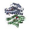
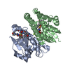
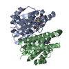
| Title | THREE-DIMENSIONAL STRUCTURE, CATALYTIC PROPERTIES AND EVOLUTION OF A SIGMA CLASS GLUTATHIONE TRANSFERASE FROM SQUID, A PROGENITOR OF THE LENS-CRYSTALLINS OF CEPHALOPODS | ||||||
 Components Components | GLUTATHIONE S-TRANSFERASE | ||||||
 Keywords Keywords | TRANSFERASE / GLUTATHIONE TRANSFERASE | ||||||
| Function / homology |  Function and homology information Function and homology informationglutathione transferase / glutathione transferase activity / glutathione metabolic process / cytoplasm Similarity search - Function | ||||||
| Biological species |  Ommastrephes sloani pacificus (Japanese flying squid) Ommastrephes sloani pacificus (Japanese flying squid) | ||||||
| Method |  X-RAY DIFFRACTION / Resolution: 2.4 Å X-RAY DIFFRACTION / Resolution: 2.4 Å | ||||||
 Authors Authors | Ji, X. / Rosenvinge, E.C.V. / Armstrong, R.N. / Gilliland, G.L. | ||||||
 Citation Citation | Journal: Biochemistry / Year: 1995 Title: Three-dimensional structure, catalytic properties, and evolution of a sigma class glutathione transferase from squid, a progenitor of the lens S-crystallins of cephalopods. Authors: Ji, X. / von Rosenvinge, E.C. / Johnson, W.W. / Tomarev, S.I. / Piatigorsky, J. / Armstrong, R.N. / Gilliland, G.L. #1: Journal: Biochemistry / Year: 1994Title: Structure and Function of the Xenobiotic Substrate Binding Site of a Glutathione S-Transferase as Revealed by X-Ray Crystallographic Analysis of Product Complexes with the Diastereomers of 9- ...Title: Structure and Function of the Xenobiotic Substrate Binding Site of a Glutathione S-Transferase as Revealed by X-Ray Crystallographic Analysis of Product Complexes with the Diastereomers of 9-(S-Glutathionyl)-10-Hydroxy-9,10-Dihydrophenanthrene Authors: Ji, X. / Sesay, M.A. / Dickert, L. / Prassad, S.M. / Johnson, W.W. / Ammon, H.L. / Armstrong, R.N. / Gilliland, G.L. #2: Journal: Biochemistry / Year: 1993Title: Snapshots Along the Reaction Coordinate of an SNAr Reaction Catalyzed by Glutathione Transferase Authors: Ji, X. / Armstrong, R.N. / Gilliland, G.L. #3: Journal: Biochemistry / Year: 1992Title: The Three-Dimensional Structure of a Glutathione S-Transferase from the Mu Gene Class. Structural Analysis of the Binary Complex of Isoenzyme 3-3 and Glutathione at 2.2 Angstroms Resolution Authors: Ji, X. / Zhang, P. / Armstrong, R.N. / Gilliland, G.L. | ||||||
| History |
|
- Structure visualization
| Structure viewer | Molecule: MolmilJmol/JSmol |
|---|
- Downloads & links
-Download
| PDBx/mmCIF format | 1gsq.cif.gz | 58 KB | Display | PDBx/mmCIF format |
|---|---|---|---|---|
| PDB format | pdb1gsq.ent.gz | 41.3 KB | Display | PDB format |
| PDBx/mmJSON format | 1gsq.json.gz | Tree view | PDBx/mmJSON format | |
| Others |  Other downloads Other downloads |
-Validation report
| Arichive directory | https://data.pdbj.org/pub/pdb/validation_reports/gs/1gsqftp://data.pdbj.org/pub/pdb/validation_reports/gs/1gsq | HTTPS FTP |
|---|
-Related structure data
| Similar structure data |
|---|


-Links
 PDBj
PDBj

- Assembly
| Deposited unit | 
| ||||||||
|---|---|---|---|---|---|---|---|---|---|
| 1 | 
| ||||||||
| Unit cell |
| ||||||||
| Atom site foot note | 1: CIS PROLINE - PRO 51 2: RESIDUE 203: ATOMS SG2, C1', C2', C3', C4', C5',C6', N2', O21, O22, N4', O41 AND O42 HAVE OCCUPANCY OF 0.70. | ||||||||
| Details | SYMMETRY THE CRYSTALLOGRAPHIC SYMMETRY TRANSFORMATIONS PRESENTED BELOW GENERATE THE SUBUNITS OF THE POLYMERIC MOLECULE. APPLIED TO RESIDUES: 1 .. 203 TO FORM THE BIOLOGICALLY ACTIVE DIMERIC MOLECULE, THE SECOND MONOMER NEEDS TO BE GENERATED BY USING THE FOLLOWING MATRIX. SYMMETRY1 1 -0.499940 0.866060 -0.000030 -0.00159 SYMMETRY2 1 0.866060 0.499940 -0.000030 0.00002 SYMMETRY3 1 -0.000010 -0.000040 -1.000000 0.00153 |
-Components
| #1: Protein | Mass: 22820.578 Da / Num. of mol.: 1 Source method: isolated from a genetically manipulated source Source: (gene. exp.) Ommastrephes sloani pacificus (Japanese flying squid)Species: Ommastrephes sloani / Strain: pacificus / Gene: CDNA INSERT OF CLONE PGST5 / Organ: DIGESTIVE GLAND / Plasmid: GST5/PET / Gene (production host): CDNA INSERT OF CLONE PGST5 / Production host:  |
|---|---|
| #2: Chemical | ChemComp-GDN / 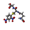  Mass: 473.415 Da / Num. of mol.: 1 / Source method: obtained synthetically / Formula: C16H19N5O10S Mass: 473.415 Da / Num. of mol.: 1 / Source method: obtained synthetically / Formula: C16H19N5O10S |
| #3: Water | ChemComp-HOH /  Mass: 18.015 Da / Num. of mol.: 154 / Source method: isolated from a natural source / Formula: H2O Mass: 18.015 Da / Num. of mol.: 154 / Source method: isolated from a natural source / Formula: H2O |
-Experimental details
-Experiment
| Experiment | Method: X-RAY DIFFRACTION |
|---|
- Sample preparation
| Crystal | Density Matthews: 3.17 Å3/Da / Density % sol: 61.16 % | |||||||||||||||||||||||||||||||||||
|---|---|---|---|---|---|---|---|---|---|---|---|---|---|---|---|---|---|---|---|---|---|---|---|---|---|---|---|---|---|---|---|---|---|---|---|---|
| Crystal | *PLUS Density % sol: 55 % | |||||||||||||||||||||||||||||||||||
| Crystal grow | *PLUS Temperature: 4 ℃ / pH: 7 / Method: vapor diffusion, sitting drop | |||||||||||||||||||||||||||||||||||
| Components of the solutions | *PLUS
|
-Data collection
| Diffraction source | Wavelength: 1.5418 |
|---|---|
| Detector | Type: ELECTRONICS COMPUTING TECHNOLOGIES / Detector: AREA DETECTOR / Date: Jan 11, 1993 |
| Radiation | Scattering type: x-ray |
| Radiation wavelength | Wavelength: 1.5418 Å / Relative weight: 1 |
| Reflection | Num. obs: 11745 / % possible obs: 99.7 % / Redundancy: 3.84 % |
| Reflection | *PLUS Highest resolution: 2.4 Å / Rmerge(I) obs: 0.087 |
- Processing
| Software |
| ||||||||||||||||||||||||||||||||||||||||||||||||||||||||||||||||||||||||||||||||||||
|---|---|---|---|---|---|---|---|---|---|---|---|---|---|---|---|---|---|---|---|---|---|---|---|---|---|---|---|---|---|---|---|---|---|---|---|---|---|---|---|---|---|---|---|---|---|---|---|---|---|---|---|---|---|---|---|---|---|---|---|---|---|---|---|---|---|---|---|---|---|---|---|---|---|---|---|---|---|---|---|---|---|---|---|---|---|
| Refinement | Resolution: 2.4→6 Å / σ(F): 4 /
| ||||||||||||||||||||||||||||||||||||||||||||||||||||||||||||||||||||||||||||||||||||
| Displacement parameters | Biso mean: 27.64 Å2 | ||||||||||||||||||||||||||||||||||||||||||||||||||||||||||||||||||||||||||||||||||||
| Refinement step | Cycle: LAST / Resolution: 2.4→6 Å
| ||||||||||||||||||||||||||||||||||||||||||||||||||||||||||||||||||||||||||||||||||||
| Refine LS restraints |
|
