 Movie
Movie Controller

[English] 日本語
 Yorodumi
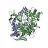
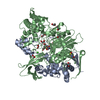
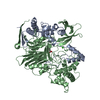
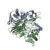
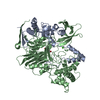
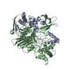
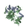
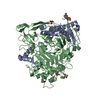
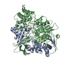
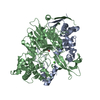
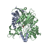
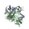
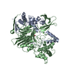
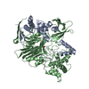
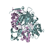
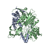
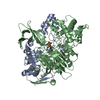
- PDB-1gm7: Crystal structures of penicillin acylase enzyme-substrate complex... -
+ Open data

- Basic information
| Entry | Database: PDB / ID: 1gm7 | ||||||
|---|---|---|---|---|---|---|---|
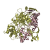
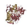
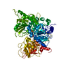
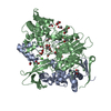
| Title | Crystal structures of penicillin acylase enzyme-substrate complexes: Structural insights into the catalytic mechanism | ||||||
 Components Components | (PENICILLIN G ACYLASE ...) x 2 | ||||||
 Keywords Keywords | HYDROLASE / ANTIBIOTIC RESISTANCE | ||||||
| Function / homology |  Function and homology information Function and homology informationpenicillin amidase activity / penicillin amidase / antibiotic biosynthetic process / periplasmic space / response to antibiotic / metal ion binding Similarity search - Function | ||||||
| Biological species |  | ||||||
| Method |  X-RAY DIFFRACTION / SYNCHROTRON / OTHER / Resolution: 1.45 Å X-RAY DIFFRACTION / SYNCHROTRON / OTHER / Resolution: 1.45 Å | ||||||
 Authors Authors | McVey, C.E. / Walsh, M.A. / Dodson, G.G. / Wilson, K.S. / Brannigan, J.A. | ||||||
 Citation Citation | Journal: J.Mol.Biol. / Year: 2001 Title: Crystal Structures of Penicillin Acylase Enzyme- Substrate Complexes: Structural Insights Into the Catalytic Mechanism Authors: Mcvey, C.E. / Walsh, M.A. / Dodson, G.G. / Wilson, K.S. / Brannigan, J.A. #1: Journal: Protein Eng. / Year: 1990 Title: Expression, Purification and Crystallisation of Penicillin G Acylase from Escherichia Coli Atcc 11105 Authors: Hunt, P.D. / Tolley, S.P. / Ward, R.J. / Hill, C.P. / Dodson, G.G. #2: Journal: Nature / Year: 1995Title: Penicillin Acylase Has a Single-Amino-Acid Catalytic Centre Authors: Duggleby, H.J. / Tolley, S.P. / Hill, C.P. / Dodson, E.J. / Dodson, G. / Moody, P.C.E. | ||||||
| History |
|
- Structure visualization
| Structure viewer | Molecule: MolmilJmol/JSmol |
|---|
- Downloads & links
-Download
| PDBx/mmCIF format | 1gm7.cif.gz | 195.7 KB | Display | PDBx/mmCIF format |
|---|---|---|---|---|
| PDB format | pdb1gm7.ent.gz | 152.8 KB | Display | PDB format |
| PDBx/mmJSON format | 1gm7.json.gz | Tree view | PDBx/mmJSON format | |
| Others |  Other downloads Other downloads |
-Validation report
| Arichive directory | https://data.pdbj.org/pub/pdb/validation_reports/gm/1gm7ftp://data.pdbj.org/pub/pdb/validation_reports/gm/1gm7 | HTTPS FTP |
|---|
-Related structure data


-Links
 PDBj
PDBj


- Assembly
| Deposited unit | 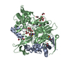
| ||||||||
|---|---|---|---|---|---|---|---|---|---|
| 1 |
| ||||||||
| Unit cell |
|
-Components
-PENICILLIN G ACYLASE ... , 2 types, 2 molecules AB
| #1: Protein | Mass: 23854.824 Da / Num. of mol.: 1 / Fragment: RESIDUES 27-235 Source method: isolated from a genetically manipulated source Source: (gene. exp.) |
|---|---|
| #2: Protein | Mass: 62386.473 Da / Num. of mol.: 1 / Fragment: RESIDUES 290-846 / Mutation: YES Source method: isolated from a genetically manipulated source Source: (gene. exp.) |
-Non-polymers , 4 types, 952 molecules 


| #3: Chemical | ChemComp-EDO /  Mass: 62.068 Da / Num. of mol.: 20 / Source method: obtained synthetically / Formula: C2H6O2 Mass: 62.068 Da / Num. of mol.: 20 / Source method: obtained synthetically / Formula: C2H6O2#4: Chemical | ChemComp-CA / |  Mass: 40.078 Da / Num. of mol.: 1 / Source method: obtained synthetically / Formula: Ca Mass: 40.078 Da / Num. of mol.: 1 / Source method: obtained synthetically / Formula: Ca#5: Chemical | ChemComp-PNN / |  Mass: 334.390 Da / Num. of mol.: 1 / Source method: obtained synthetically / Formula: C16H18N2O4S / Comment: antibiotic*YM Mass: 334.390 Da / Num. of mol.: 1 / Source method: obtained synthetically / Formula: C16H18N2O4S / Comment: antibiotic*YM#6: Water | ChemComp-HOH / | Mass: 18.015 Da / Num. of mol.: 930 / Source method: isolated from a natural source / Formula: H2O |
|---|
-Details
| Has protein modification | Y |
|---|
-Experimental details
-Experiment
| Experiment | Method: X-RAY DIFFRACTION / Number of used crystals: 1 |
|---|
- Sample preparation
| Crystal | Density Matthews: 2.43 Å3/Da / Density % sol: 42.7 % |
|---|---|
| Crystal grow | pH: 7.5 / Details: pH 7.50 |
-Data collection
| Diffraction | Mean temperature: 100 K |
|---|---|
| Diffraction source | Source: SYNCHROTRON / Site: EMBL/DESY, HAMBURG  / Beamline: BW7B / Wavelength: 0.89 / Beamline: BW7B / Wavelength: 0.89 |
| Detector | Type: MAR scanner 300 mm plate / Detector: IMAGE PLATE |
| Radiation | Protocol: SINGLE WAVELENGTH / Monochromatic (M) / Laue (L): M / Scattering type: x-ray |
| Radiation wavelength | Wavelength: 0.89 Å / Relative weight: 1 |
| Reflection | Resolution: 1.45→30 Å / Num. obs: 144035 / % possible obs: 99.6 % / Redundancy: 3.4 % / Biso Wilson estimate: 13.4 Å2 / Rmerge(I) obs: 0.061 / Net I/σ(I): 18.7 |
| Reflection shell | Resolution: 1.45→1.47 Å / Rmerge(I) obs: 0.339 / Mean I/σ(I) obs: 3.3 / % possible all: 99.9 |
- Processing
| Software |
| ||||||||||||||||||||||||||||||||||||||||||||||||||||||||||||||||||||||||||||||||||||
|---|---|---|---|---|---|---|---|---|---|---|---|---|---|---|---|---|---|---|---|---|---|---|---|---|---|---|---|---|---|---|---|---|---|---|---|---|---|---|---|---|---|---|---|---|---|---|---|---|---|---|---|---|---|---|---|---|---|---|---|---|---|---|---|---|---|---|---|---|---|---|---|---|---|---|---|---|---|---|---|---|---|---|---|---|---|
| Refinement | Method to determine structure: OTHER / Resolution: 1.45→30 Å / SU B: 0.96 / SU ML: 0.036 / Cross valid method: THROUGHOUT / σ(F): 0 / ESU R: 0.055 / ESU R Free: 0.058
| ||||||||||||||||||||||||||||||||||||||||||||||||||||||||||||||||||||||||||||||||||||
| Displacement parameters | Biso mean: 14.8 Å2 | ||||||||||||||||||||||||||||||||||||||||||||||||||||||||||||||||||||||||||||||||||||
| Refinement step | Cycle: LAST / Resolution: 1.45→30 Å
| ||||||||||||||||||||||||||||||||||||||||||||||||||||||||||||||||||||||||||||||||||||
| Refine LS restraints |
|
