 Movie
Movie Controller

[English] 日本語
 Yorodumi
- PDB-1e3a: A slow processing precursor penicillin acylase from Escherichia coli -
+ Open data

- Basic information
| Entry | Database: PDB / ID: 1e3a | ||||||
|---|---|---|---|---|---|---|---|
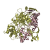
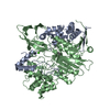
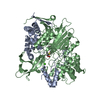
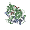
| Title | A slow processing precursor penicillin acylase from Escherichia coli | ||||||
 Components Components | (PENICILLIN AMIDASE ...) x 2 | ||||||
 Keywords Keywords | ANTIBIOTIC RESISTANCE / AMIDASE / NTN-HYDROLASE / HYDROLYSIS OF PENICILLIN G ACYLASE | ||||||
| Function / homology |  Function and homology information Function and homology informationpenicillin amidase activity / penicillin amidase / antibiotic biosynthetic process / periplasmic space / response to antibiotic / metal ion binding Similarity search - Function | ||||||
| Biological species |  | ||||||
| Method |  X-RAY DIFFRACTION / SYNCHROTRON / MOLECULAR REPLACEMENT / Resolution: 1.8 Å X-RAY DIFFRACTION / SYNCHROTRON / MOLECULAR REPLACEMENT / Resolution: 1.8 Å | ||||||
 Authors Authors | Hewitt, L. / Kasche, V. / Lummer, K. / Lewis, R.J. / Murshudov, G.N. / Verma, C.S. / Dodson, G.G. / Wilson, K.S. | ||||||
 Citation Citation | Journal: J.Mol.Biol. / Year: 2000 Title: Structure of a Slow Processing Precursor Penicillin Acylase from Escherichia Coli Reveals the Linker Peptide Blocking the Active-Site Cleft Authors: Hewitt, L. / Kasche, V. / Lummer, K. / Lewis, R.J. / Murshudov, G.N. / Verma, C.S. / Dodson, G.G. / Wilson, K.S. #1: Journal: Acta Crystallogr.,Sect.D / Year: 1999 Title: Crystallisation of a Precursor Penicillin Acylase from Escherichia Coli Authors: Hewitt, L. / Kasche, V. / Lummer, K. / Rieks, A. / Wilson, K.S. | ||||||
| History |
|
- Structure visualization
| Structure viewer | Molecule: MolmilJmol/JSmol |
|---|
- Downloads & links
-Download
| PDBx/mmCIF format | 1e3a.cif.gz | 202.8 KB | Display | PDBx/mmCIF format |
|---|---|---|---|---|
| PDB format | pdb1e3a.ent.gz | 156.8 KB | Display | PDB format |
| PDBx/mmJSON format | 1e3a.json.gz | Tree view | PDBx/mmJSON format | |
| Others |  Other downloads Other downloads |
-Validation report
| Arichive directory | https://data.pdbj.org/pub/pdb/validation_reports/e3/1e3aftp://data.pdbj.org/pub/pdb/validation_reports/e3/1e3a | HTTPS FTP |
|---|
-Related structure data
| Related structure data |  1pnkS S: Starting model for refinement |
|---|---|
| Similar structure data |


-Links
 PDBj
PDBj


- Assembly
| Deposited unit | 
| ||||||||
|---|---|---|---|---|---|---|---|---|---|
| 1 |
| ||||||||
| Unit cell |
|
-Components
-PENICILLIN AMIDASE ... , 2 types, 2 molecules AB
| #1: Protein | Mass: 28963.695 Da / Num. of mol.: 1 / Fragment: PENICILLIN AMIDASE RESIDUES 29-286 Source method: isolated from a genetically manipulated source Source: (gene. exp.) |
|---|---|
| #2: Protein | Mass: 62684.770 Da / Num. of mol.: 1 / Fragment: PENICILLIN AMIDASE RESIDUES 287-846 / Mutation: YES Source method: isolated from a genetically manipulated source Source: (gene. exp.) |
-Non-polymers , 4 types, 1198 molecules 


| #3: Chemical | ChemComp-CA /  Mass: 40.078 Da / Num. of mol.: 1 / Source method: obtained synthetically / Formula: Ca Mass: 40.078 Da / Num. of mol.: 1 / Source method: obtained synthetically / Formula: Ca | ||
|---|---|---|---|
| #4: Chemical | ChemComp-CL /  Mass: 35.453 Da / Num. of mol.: 1 / Source method: obtained synthetically / Formula: Cl Mass: 35.453 Da / Num. of mol.: 1 / Source method: obtained synthetically / Formula: Cl | ||
| #5: Chemical |  Mass: 62.068 Da / Num. of mol.: 3 / Source method: obtained synthetically / Formula: C2H6O2 Mass: 62.068 Da / Num. of mol.: 3 / Source method: obtained synthetically / Formula: C2H6O2#6: Water | ChemComp-HOH / | Mass: 18.015 Da / Num. of mol.: 1193 / Source method: isolated from a natural source / Formula: H2O |
-Details
| Compound details | CHAIN B ENGINEERED| Sequence details | SWISS-PROT SEQ: SIGNAL REMOVED ON TRANSLOCAT | |
|---|
-Experimental details
-Experiment
| Experiment | Method: X-RAY DIFFRACTION / Number of used crystals: 1 |
|---|
- Sample preparation
| Crystal | Density Matthews: 2.17 Å3/Da / Density % sol: 43 % | |||||||||||||||||||||||||||||||||||
|---|---|---|---|---|---|---|---|---|---|---|---|---|---|---|---|---|---|---|---|---|---|---|---|---|---|---|---|---|---|---|---|---|---|---|---|---|
| Crystal grow | pH: 7.2 Details: 50MM MOPS PH7.2, 18-20% PEG 5KME, 10MM CACL2, pH 7.20 | |||||||||||||||||||||||||||||||||||
| Crystal | *PLUS Density % sol: 43 % | |||||||||||||||||||||||||||||||||||
| Crystal grow | *PLUS Temperature: 18 ℃ / Method: vapor diffusion, hanging drop / pH: 7.2 | |||||||||||||||||||||||||||||||||||
| Components of the solutions | *PLUS
|
-Data collection
| Diffraction | Mean temperature: 120 K |
|---|---|
| Diffraction source | Source: SYNCHROTRON / Site: EMBL/DESY, HAMBURG  / Beamline: X11 / Wavelength: 0.9096 / Beamline: X11 / Wavelength: 0.9096 |
| Detector | Type: MARRESEARCH / Detector: IMAGE PLATE / Date: Nov 15, 1997 / Details: SEGMENTED MIRROR |
| Radiation | Monochromator: SI(111) / Protocol: SINGLE WAVELENGTH / Monochromatic (M) / Laue (L): M / Scattering type: x-ray |
| Radiation wavelength | Wavelength: 0.9096 Å / Relative weight: 1 |
| Reflection | Resolution: 1.8→20 Å / Num. obs: 70642 / % possible obs: 96.9 % / Redundancy: 1.9 % / Biso Wilson estimate: 12.2 Å2 / Rmerge(I) obs: 0.063 / Net I/σ(I): 10.3 |
| Reflection shell | Resolution: 1.8→1.86 Å / Redundancy: 1.5 % / Rmerge(I) obs: 0.216 / Mean I/σ(I) obs: 3.16 / % possible all: 89.1 |
| Reflection | *PLUS Num. measured all: 133678 |
| Reflection shell | *PLUS % possible obs: 89.1 % |
- Processing
| Software |
| ||||||||||||||||||||||||||||||||||||||||||||||||||||||||||||||||||||||||||||||||||||
|---|---|---|---|---|---|---|---|---|---|---|---|---|---|---|---|---|---|---|---|---|---|---|---|---|---|---|---|---|---|---|---|---|---|---|---|---|---|---|---|---|---|---|---|---|---|---|---|---|---|---|---|---|---|---|---|---|---|---|---|---|---|---|---|---|---|---|---|---|---|---|---|---|---|---|---|---|---|---|---|---|---|---|---|---|---|
| Refinement | Method to determine structure: MOLECULAR REPLACEMENT Starting model: PDB ENTRY 1PNK Resolution: 1.8→20 Å / SU B: 2.5 / SU ML: 0.08 / Cross valid method: THROUGHOUT / σ(F): 0 / ESU R: 0.13 / ESU R Free: 0.12
| ||||||||||||||||||||||||||||||||||||||||||||||||||||||||||||||||||||||||||||||||||||
| Displacement parameters | Biso mean: 15.1 Å2 | ||||||||||||||||||||||||||||||||||||||||||||||||||||||||||||||||||||||||||||||||||||
| Refinement step | Cycle: LAST / Resolution: 1.8→20 Å
| ||||||||||||||||||||||||||||||||||||||||||||||||||||||||||||||||||||||||||||||||||||
| Refine LS restraints |
| ||||||||||||||||||||||||||||||||||||||||||||||||||||||||||||||||||||||||||||||||||||
| Software | *PLUS Name: REFMAC / Classification: refinement | ||||||||||||||||||||||||||||||||||||||||||||||||||||||||||||||||||||||||||||||||||||
| Refinement | *PLUS Lowest resolution: 20 Å | ||||||||||||||||||||||||||||||||||||||||||||||||||||||||||||||||||||||||||||||||||||
| Solvent computation | *PLUS | ||||||||||||||||||||||||||||||||||||||||||||||||||||||||||||||||||||||||||||||||||||
| Displacement parameters | *PLUS | ||||||||||||||||||||||||||||||||||||||||||||||||||||||||||||||||||||||||||||||||||||
| Refine LS restraints | *PLUS Type: p_bond_d / Dev ideal: 0.009 |
