 Movie
Movie Controller


+ Open data

- Basic information
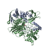
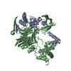
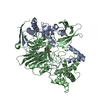
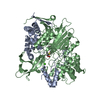
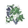
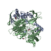
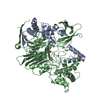
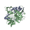
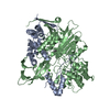
| Entry | Database: PDB / ID: 1ai5 | ||||||
|---|---|---|---|---|---|---|---|
| Title | PENICILLIN ACYLASE COMPLEXED WITH M-NITROPHENYLACETIC ACID | ||||||
 Components Components | (PENICILLIN AMIDOHYDROLASE) x 2 | ||||||
 Keywords Keywords | ANTIBIOTIC RESISTANCE / LIGAND INDUCED CONFORMATIONAL CHANGE / HYDROLASE | ||||||
| Function / homology |  Function and homology information Function and homology informationpenicillin amidase activity / penicillin amidase / antibiotic biosynthetic process / periplasmic space / response to antibiotic / metal ion binding Similarity search - Function | ||||||
| Biological species |  | ||||||
| Method |  X-RAY DIFFRACTION / SYNCHROTRON / STRUCTURE ISOMORPHOUS TO NATIVE / Resolution: 2.36 Å X-RAY DIFFRACTION / SYNCHROTRON / STRUCTURE ISOMORPHOUS TO NATIVE / Resolution: 2.36 Å | ||||||
 Authors Authors | Done, S.H. | ||||||
 Citation Citation | Journal: J.Mol.Biol. / Year: 1998 Title: Ligand-induced conformational change in penicillin acylase. Authors: Done, S.H. / Brannigan, J.A. / Moody, P.C.E. / Hubbard, R.E. #2: Journal: Nature / Year: 1995Title: Penicillin Acylase Has a Single-Amino-Acid Catalytic Centre Authors: Duggleby, H.J. / Tolley, S.P. / Hill, C.P. / Dodson, E.J. / Dodson, G. / Moody, P.C. #3: Journal: Protein Eng. / Year: 1990Title: Expression, Purification and Crystallization of Penicillin G Acylase from Escherichia Coli Atcc 11105 Authors: Hunt, P.D. / Tolley, S.P. / Ward, R.J. / Hill, C.P. / Dodson, G.G. | ||||||
| History |
|
- Structure visualization
| Structure viewer | Molecule: MolmilJmol/JSmol |
|---|
- Downloads & links
-Download
| PDBx/mmCIF format | 1ai5.cif.gz | 180.3 KB | Display | PDBx/mmCIF format |
|---|---|---|---|---|
| PDB format | pdb1ai5.ent.gz | 139.3 KB | Display | PDB format |
| PDBx/mmJSON format | 1ai5.json.gz | Tree view | PDBx/mmJSON format | |
| Others |  Other downloads Other downloads |
-Validation report
| Arichive directory | https://data.pdbj.org/pub/pdb/validation_reports/ai/1ai5ftp://data.pdbj.org/pub/pdb/validation_reports/ai/1ai5 | HTTPS FTP |
|---|
-Related structure data
| Related structure data |  1ai4C  1ai6C  1ai7C 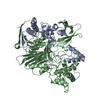 1ajnC  1ajpC 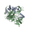 1ajqC C: citing same article ( |
|---|---|
| Similar structure data |

-Links
 PDBj
PDBj


- Assembly
| Deposited unit | 
| ||||||||
|---|---|---|---|---|---|---|---|---|---|
| 1 |
| ||||||||
| Unit cell |
|
-Components
| #1: Protein | Mass: 23838.824 Da / Num. of mol.: 1 / Source method: isolated from a natural source / Source: (natural) |
|---|---|
| #2: Protein | Mass: 62428.512 Da / Num. of mol.: 1 / Source method: isolated from a natural source / Source: (natural) |
| #3: Chemical | ChemComp-CA /   Mass: 40.078 Da / Num. of mol.: 1 / Source method: obtained synthetically / Formula: Ca Mass: 40.078 Da / Num. of mol.: 1 / Source method: obtained synthetically / Formula: Ca |
| #4: Chemical | ChemComp-MNP / 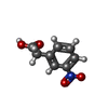  Mass: 181.145 Da / Num. of mol.: 1 / Source method: obtained synthetically / Formula: C8H7NO4 Mass: 181.145 Da / Num. of mol.: 1 / Source method: obtained synthetically / Formula: C8H7NO4 |
| #5: Water | ChemComp-HOH /  Mass: 18.015 Da / Num. of mol.: 663 / Source method: isolated from a natural source / Formula: H2O Mass: 18.015 Da / Num. of mol.: 663 / Source method: isolated from a natural source / Formula: H2O |
-Experimental details
-Experiment
| Experiment | Method: X-RAY DIFFRACTION / Number of used crystals: 1 |
|---|
- Sample preparation
| Crystal | Density Matthews: 3.06 Å3/Da / Density % sol: 59.5 % | ||||||||||||||||||||||||||||||
|---|---|---|---|---|---|---|---|---|---|---|---|---|---|---|---|---|---|---|---|---|---|---|---|---|---|---|---|---|---|---|---|
| Crystal grow | Method: streak seeded / pH: 7.2 Details: CRYSTALLIZED FROM 10% PEG 8000, 50MM MOPS, PH 7.2, STREAK SEEDED. SOAKED IN 5MM M-NITROPHENYLACETIC ACID, streak seeded | ||||||||||||||||||||||||||||||
| Crystal grow | *PLUS Method: vapor diffusion, hanging drop | ||||||||||||||||||||||||||||||
| Components of the solutions | *PLUS
|
-Data collection
| Diffraction | Mean temperature: 300 K |
|---|---|
| Diffraction source | Source: SYNCHROTRON / Site: EMBL/DESY, HAMBURG  / Beamline: X31 / Wavelength: 0.98 / Beamline: X31 / Wavelength: 0.98 |
| Detector | Type: MARRESEARCH / Detector: IMAGE PLATE / Date: Aug 1, 1995 |
| Radiation | Monochromatic (M) / Laue (L): M / Scattering type: x-ray |
| Radiation wavelength | Wavelength: 0.98 Å / Relative weight: 1 |
| Reflection | Resolution: 2.36→26.12 Å / Num. obs: 34287 / % possible obs: 97.4 % / Redundancy: 2.1 % / Biso Wilson estimate: 19.02 Å2 / Rmerge(I) obs: 0.091 / Net I/σ(I): 8.2 |
| Reflection shell | Resolution: 2.35→2.48 Å / Redundancy: 2 % / Rmerge(I) obs: 0.331 / Mean I/σ(I) obs: 2.3 / % possible all: 93.4 |
| Reflection | *PLUS Num. measured all: 71097 |
- Processing
| Software |
| ||||||||||||||||||||||||||||||||||||||||||||||||||||||||||||||||||||||||||||||||||||
|---|---|---|---|---|---|---|---|---|---|---|---|---|---|---|---|---|---|---|---|---|---|---|---|---|---|---|---|---|---|---|---|---|---|---|---|---|---|---|---|---|---|---|---|---|---|---|---|---|---|---|---|---|---|---|---|---|---|---|---|---|---|---|---|---|---|---|---|---|---|---|---|---|---|---|---|---|---|---|---|---|---|---|---|---|---|
| Refinement | Method to determine structure: STRUCTURE ISOMORPHOUS TO NATIVE Resolution: 2.36→26.12 Å / Cross valid method: FREE_R / σ(F): 0
| ||||||||||||||||||||||||||||||||||||||||||||||||||||||||||||||||||||||||||||||||||||
| Displacement parameters | Biso mean: 30.47 Å2 | ||||||||||||||||||||||||||||||||||||||||||||||||||||||||||||||||||||||||||||||||||||
| Refinement step | Cycle: LAST / Resolution: 2.36→26.12 Å
| ||||||||||||||||||||||||||||||||||||||||||||||||||||||||||||||||||||||||||||||||||||
| Refine LS restraints |
| ||||||||||||||||||||||||||||||||||||||||||||||||||||||||||||||||||||||||||||||||||||
| Software | *PLUS Name: REFMAC / Classification: refinement | ||||||||||||||||||||||||||||||||||||||||||||||||||||||||||||||||||||||||||||||||||||
| Refinement | *PLUS Num. reflection all: 34287 / Rfactor all: 0.1493 | ||||||||||||||||||||||||||||||||||||||||||||||||||||||||||||||||||||||||||||||||||||
| Solvent computation | *PLUS | ||||||||||||||||||||||||||||||||||||||||||||||||||||||||||||||||||||||||||||||||||||
| Displacement parameters | *PLUS |
