 Movie
Movie Controller


+ Open data

- Basic information
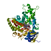
| Entry | Database: PDB / ID: 1gl4 | ||||||
|---|---|---|---|---|---|---|---|
| Title | Nidogen-1 G2/Perlecan IG3 Complex | ||||||
 Components Components |
| ||||||
 Keywords Keywords | BASEMENT MEMBRANE / IMMUNOGLOBULIN-LIKE DOMAIN / EXTRACELLULAR MATRIX / PROTEOGLYCAN / HEPARAN SULFATE | ||||||
| Function / homology |  Function and homology information Function and homology informationLaminin interactions / gel phase of basement membrane / elastic fiber / collagen V binding / regulation of basement membrane organization / glomerular basement membrane development / microfibril / laminin-1 binding / negative regulation of synaptic transmission, cholinergic / positive regulation of integrin-mediated signaling pathway ...Laminin interactions / gel phase of basement membrane / elastic fiber / collagen V binding / regulation of basement membrane organization / glomerular basement membrane development / microfibril / laminin-1 binding / negative regulation of synaptic transmission, cholinergic / positive regulation of integrin-mediated signaling pathway / Degradation of the extracellular matrix / cartilage development involved in endochondral bone morphogenesis / tissue development / embryonic skeletal system morphogenesis / dystroglycan binding / endochondral ossification / protein complex involved in cell-matrix adhesion / protein localization to synapse / negative regulation of cell adhesion / positive regulation of cell-substrate adhesion / negative regulation of amyloid fibril formation / extracellular matrix binding / proteoglycan binding / cardiac muscle tissue development / positive regulation of muscle cell differentiation / smoothened signaling pathway / basement membrane / chondrocyte differentiation / collagen binding / positive regulation of endothelial cell proliferation / extracellular matrix organization / laminin binding / positive regulation of cell adhesion / receptor-mediated endocytosis / animal organ morphogenesis / cell-matrix adhesion / cell periphery / brain development / Golgi lumen / intracellular protein localization / extracellular matrix / protease binding / angiogenesis / molecular adaptor activity / negative regulation of neuron apoptotic process / negative regulation of cell population proliferation / calcium ion binding / extracellular region Similarity search - Function | ||||||
| Biological species |  | ||||||
| Method |  X-RAY DIFFRACTION / SYNCHROTRON / MOLECULAR REPLACEMENT / Resolution: 2 Å X-RAY DIFFRACTION / SYNCHROTRON / MOLECULAR REPLACEMENT / Resolution: 2 Å | ||||||
 Authors Authors | Kvansakul, M. / Hopf, M. / Ries, A. / Timpl, R. / Hohenester, E. | ||||||
 Citation Citation | Journal: Embo J. / Year: 2001 Title: Structural Basis for the High-Affinity Interaction of Nidogen-1 with Immunoglobulin-Like Domain 3 of Perlecan Authors: Kvansakul, M. / Hopf, M. / Ries, A. / Timpl, R. / Hohenester, E. #1: Journal: Nat.Struct.Biol . / Year: 2001Title: Crystal Structure and Mutational Analysis of a Perlecan-Binding Fragment of Nidogen-1 Authors: Hopf, M. / Gohring, W. / Ries, A. / Timpl, R. / Hohenester, E. #2: Journal: J.Mol.Biol. / Year: 2001 Title: Mapping of Binding Sites for Nidogens, Fibulin-2, Fibronectin and Heparin to Different Ig Modules of Perlecan Authors: Hopf, M. / Gohring, W. / Mann, K. / Timpl, R. | ||||||
| History |
| ||||||
| Remark 700 | SHEET THE SHEET STRUCTURE OF THIS MOLECULE IS BIFURCATED. IN ORDER TO REPRESENT THIS FEATURE IN ... SHEET THE SHEET STRUCTURE OF THIS MOLECULE IS BIFURCATED. IN ORDER TO REPRESENT THIS FEATURE IN THE SHEET RECORDS BELOW, TWO SHEETS ARE DEFINED. |
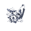
- Structure visualization
| Structure viewer | Molecule: MolmilJmol/JSmol |
|---|
- Downloads & links
-Download
| PDBx/mmCIF format | 1gl4.cif.gz | 88.1 KB | Display | PDBx/mmCIF format |
|---|---|---|---|---|
| PDB format | pdb1gl4.ent.gz | 65.1 KB | Display | PDB format |
| PDBx/mmJSON format | 1gl4.json.gz | Tree view | PDBx/mmJSON format | |
| Others |  Other downloads Other downloads |
-Validation report
| Arichive directory | https://data.pdbj.org/pub/pdb/validation_reports/gl/1gl4ftp://data.pdbj.org/pub/pdb/validation_reports/gl/1gl4 | HTTPS FTP |
|---|
-Related structure data
| Related structure data |  1h4uS S: Starting model for refinement |
|---|---|
| Similar structure data |


-Links
 PDBj
PDBj


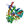
- Assembly
| Deposited unit | 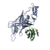
| ||||||||
|---|---|---|---|---|---|---|---|---|---|
| 1 |
| ||||||||
| Unit cell |
|
-Components
| #1: Protein | Mass: 31350.930 Da / Num. of mol.: 1 / Fragment: G2 RESIDUES 385-665 Source method: isolated from a genetically manipulated source Source: (gene. exp.)  HOMO SAPIENS (human) / References: UniProt: P10493 HOMO SAPIENS (human) / References: UniProt: P10493 |
|---|---|
| #2: Protein | Mass: 10562.085 Da / Num. of mol.: 1 / Fragment: IG3 RESIDUES 1765-1858 Source method: isolated from a genetically manipulated source Details: VECTOR-DERIVED N-TERMINAL APLA / Source: (gene. exp.) HOMO SAPIENS (human) / References: UniProt: Q05793 |
| #3: Chemical | ChemComp-ZN /   Mass: 65.409 Da / Num. of mol.: 1 / Source method: obtained synthetically / Formula: Zn Mass: 65.409 Da / Num. of mol.: 1 / Source method: obtained synthetically / Formula: Zn |
| #4: Chemical | ChemComp-EPE /   Mass: 238.305 Da / Num. of mol.: 1 / Source method: obtained synthetically / Formula: C8H18N2O4S / Comment: pH buffer*YM Mass: 238.305 Da / Num. of mol.: 1 / Source method: obtained synthetically / Formula: C8H18N2O4S / Comment: pH buffer*YM |
| #5: Water | ChemComp-HOH /  Mass: 18.015 Da / Num. of mol.: 174 / Source method: isolated from a natural source / Formula: H2O Mass: 18.015 Da / Num. of mol.: 174 / Source method: isolated from a natural source / Formula: H2O |
| Has protein modification | Y |
-Experimental details
-Experiment
| Experiment | Method: X-RAY DIFFRACTION / Number of used crystals: 1 |
|---|
- Sample preparation
| Crystal | Density Matthews: 2.46 Å3/Da / Density % sol: 50.1 % | ||||||||||||||||||||||||||||||||||||
|---|---|---|---|---|---|---|---|---|---|---|---|---|---|---|---|---|---|---|---|---|---|---|---|---|---|---|---|---|---|---|---|---|---|---|---|---|---|
| Crystal grow | pH: 7.5 / Details: 10% PEG 6000, 5% MPD, 0.1 M HEPES PH 7.5 | ||||||||||||||||||||||||||||||||||||
| Crystal grow | *PLUS Method: vapor diffusion, hanging drop | ||||||||||||||||||||||||||||||||||||
| Components of the solutions | *PLUS
|
-Data collection
| Diffraction | Mean temperature: 100 K |
|---|---|
| Diffraction source | Source: SYNCHROTRON / Site: SRS  / Beamline: PX9.6 / Wavelength: 0.87 / Beamline: PX9.6 / Wavelength: 0.87 |
| Detector | Type: QUANTUM4 / Detector: CCD / Date: Nov 6, 2000 |
| Radiation | Protocol: SINGLE WAVELENGTH / Monochromatic (M) / Laue (L): M / Scattering type: x-ray |
| Radiation wavelength | Wavelength: 0.87 Å / Relative weight: 1 |
| Reflection | Resolution: 2→20 Å / Num. obs: 29096 / % possible obs: 99.8 % / Observed criterion σ(I): 0 / Redundancy: 3.7 % / Biso Wilson estimate: 18.3 Å2 / Rmerge(I) obs: 0.089 / Net I/σ(I): 11.4 |
| Reflection shell | Resolution: 2→2.11 Å / Redundancy: 3.7 % / Rmerge(I) obs: 0.256 / Mean I/σ(I) obs: 4.7 / % possible all: 99.8 |
| Reflection | *PLUS Highest resolution: 2 Å / Lowest resolution: 20 Å |
- Processing
| Software |
| ||||||||||||||||||||||||||||||||||||||||||||||||||||||||||||||||||||||||||||||||
|---|---|---|---|---|---|---|---|---|---|---|---|---|---|---|---|---|---|---|---|---|---|---|---|---|---|---|---|---|---|---|---|---|---|---|---|---|---|---|---|---|---|---|---|---|---|---|---|---|---|---|---|---|---|---|---|---|---|---|---|---|---|---|---|---|---|---|---|---|---|---|---|---|---|---|---|---|---|---|---|---|---|
| Refinement | Method to determine structure: MOLECULAR REPLACEMENT Starting model: 1H4U Resolution: 2→20 Å / Data cutoff high absF: 10000 / Isotropic thermal model: INDIVIDUAL RESTRAINED / Cross valid method: THROUGHOUT / σ(F): 0
| ||||||||||||||||||||||||||||||||||||||||||||||||||||||||||||||||||||||||||||||||
| Refinement step | Cycle: LAST / Resolution: 2→20 Å
| ||||||||||||||||||||||||||||||||||||||||||||||||||||||||||||||||||||||||||||||||
| Refine LS restraints |
| ||||||||||||||||||||||||||||||||||||||||||||||||||||||||||||||||||||||||||||||||
| LS refinement shell | Resolution: 2→2.01 Å / Total num. of bins used: 50
| ||||||||||||||||||||||||||||||||||||||||||||||||||||||||||||||||||||||||||||||||
| Xplor file |
| ||||||||||||||||||||||||||||||||||||||||||||||||||||||||||||||||||||||||||||||||
| Refinement | *PLUS Highest resolution: 2 Å / Lowest resolution: 20 Å / Rfactor obs: 0.21 / Rfactor Rfree: 0.245 / Rfactor Rwork: 0.213 | ||||||||||||||||||||||||||||||||||||||||||||||||||||||||||||||||||||||||||||||||
| Solvent computation | *PLUS | ||||||||||||||||||||||||||||||||||||||||||||||||||||||||||||||||||||||||||||||||
| Displacement parameters | *PLUS | ||||||||||||||||||||||||||||||||||||||||||||||||||||||||||||||||||||||||||||||||
| Refine LS restraints | *PLUS Type: c_angle_deg / Dev ideal: 1.2 |
