 Movie
Movie Controller


+ Open data

- Basic information
| Entry | Database: PDB / ID: 7ck3 | ||||||
|---|---|---|---|---|---|---|---|
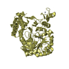
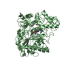
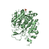
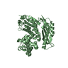
| Title | Crystal structure of Arabidopsis CESA3 catalytic domain | ||||||
 Components Components | Cellulose synthase A catalytic subunit 3 [UDP-forming],Cellulose synthase A catalytic subunit 3 [UDP-forming] | ||||||
 Keywords Keywords | PLANT PROTEIN / enzyme / synthase | ||||||
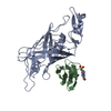
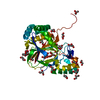
| Function / homology |  Function and homology information Function and homology informationplant-type secondary cell wall biogenesis / plant-type primary cell wall biogenesis / cellulose synthase activity / cellulose synthase (UDP-forming) / cellulose synthase (UDP-forming) activity / cellulose biosynthetic process / plasmodesma / trans-Golgi network / defense response / cell wall organization ...plant-type secondary cell wall biogenesis / plant-type primary cell wall biogenesis / cellulose synthase activity / cellulose synthase (UDP-forming) / cellulose synthase (UDP-forming) activity / cellulose biosynthetic process / plasmodesma / trans-Golgi network / defense response / cell wall organization / endosome / Golgi membrane / Golgi apparatus / protein homodimerization activity / zinc ion binding / plasma membrane Similarity search - Function | ||||||
| Biological species |  | ||||||
| Method |  X-RAY DIFFRACTION / SYNCHROTRON / MOLECULAR REPLACEMENT / Resolution: 2.9 Å X-RAY DIFFRACTION / SYNCHROTRON / MOLECULAR REPLACEMENT / Resolution: 2.9 Å | ||||||
 Authors Authors | Qiao, Z. / Gao, Y.G. | ||||||
| Funding support |  Singapore, 1items Singapore, 1items
| ||||||
 Citation Citation | Journal: Proc.Natl.Acad.Sci.USA / Year: 2021 Title: Structure of Arabidopsis CESA3 catalytic domain with its substrate UDP-glucose provides insight into the mechanism of cellulose synthesis. Authors: Qiao, Z. / Lampugnani, E.R. / Yan, X.F. / Khan, G.A. / Saw, W.G. / Hannah, P. / Qian, F. / Calabria, J. / Miao, Y. / Gruber, G. / Persson, S. / Gao, Y.G. | ||||||
| History |
|
- Structure visualization
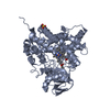
| Structure viewer | Molecule: MolmilJmol/JSmol |
|---|
- Downloads & links
-Download
| PDBx/mmCIF format | 7ck3.cif.gz | 353.4 KB | Display | PDBx/mmCIF format |
|---|---|---|---|---|
| PDB format | pdb7ck3.ent.gz | 252.6 KB | Display | PDB format |
| PDBx/mmJSON format | 7ck3.json.gz | Tree view | PDBx/mmJSON format | |
| Others |  Other downloads Other downloads |
-Validation report
| Arichive directory | https://data.pdbj.org/pub/pdb/validation_reports/ck/7ck3ftp://data.pdbj.org/pub/pdb/validation_reports/ck/7ck3 | HTTPS FTP |
|---|
-Related structure data
| Related structure data |  7ck1SC  7ck2C S: Starting model for refinement C: citing same article ( |
|---|---|
| Similar structure data |


-Links
 PDBj
PDBj

- Assembly
| Deposited unit | 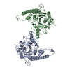
| ||||||||||||||||||||||||||||||
|---|---|---|---|---|---|---|---|---|---|---|---|---|---|---|---|---|---|---|---|---|---|---|---|---|---|---|---|---|---|---|---|
| 1 | 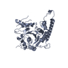
| ||||||||||||||||||||||||||||||
| 2 | 
| ||||||||||||||||||||||||||||||
| Unit cell |
| ||||||||||||||||||||||||||||||
| Noncrystallographic symmetry (NCS) | NCS domain:
NCS domain segments:
NCS oper: (Code: givenMatrix: (0.049231147085, 0.971094932843, -0.233561395704), (0.969769025593, -0.102433902371, -0.221484384652), (-0.239006968844, -0.215596676811, -0.946780725296)Vector: -8. ...NCS oper: (Code: given Matrix: (0.049231147085, 0.971094932843, -0.233561395704), Vector: |
-Components
| #1: Protein | Mass: 48078.688 Da / Num. of mol.: 2 Source method: isolated from a genetically manipulated source Source: (gene. exp.) Gene: CESA3, ATHB, CEV1, ELI1, IXR1, RSW5, At5g05170, K2A11.4 Production host:  References: UniProt: Q941L0, cellulose synthase (UDP-forming) #2: Water | ChemComp-HOH / |  Mass: 18.015 Da / Num. of mol.: 1 / Source method: isolated from a natural source / Formula: H2O Mass: 18.015 Da / Num. of mol.: 1 / Source method: isolated from a natural source / Formula: H2OHas protein modification | Y | |
|---|
-Experimental details
-Experiment
| Experiment | Method: X-RAY DIFFRACTION / Number of used crystals: 1 |
|---|
- Sample preparation
| Crystal | Density Matthews: 2.4 Å3/Da / Density % sol: 48.81 % |
|---|---|
| Crystal grow | Temperature: 291 K / Method: evaporation / Details: PEG3350 |
-Data collection
| Diffraction | Mean temperature: 80 K / Serial crystal experiment: N |
|---|---|
| Diffraction source | Source: SYNCHROTRON / Site: Australian Synchrotron  / Beamline: MX2 / Wavelength: 0.9537 Å / Beamline: MX2 / Wavelength: 0.9537 Å |
| Detector | Type: DECTRIS EIGER X 16M / Detector: PIXEL / Date: Aug 11, 2018 |
| Radiation | Protocol: SINGLE WAVELENGTH / Monochromatic (M) / Laue (L): M / Scattering type: x-ray |
| Radiation wavelength | Wavelength: 0.9537 Å / Relative weight: 1 |
| Reflection | Resolution: 2.9→40 Å / Num. obs: 21506 / % possible obs: 100 % / Redundancy: 41.19 % / Biso Wilson estimate: 66.64 Å2 / CC1/2: 0.99 / Net I/σ(I): 6.55 |
| Reflection shell | Resolution: 2.9→2.98 Å / Mean I/σ(I) obs: 0.95 / Num. unique obs: 1457 / CC1/2: 0.42 / % possible all: 99.9 |
- Processing
| Software |
| |||||||||||||||||||||||||||||||||||||||||||||||||||||||||||||||
|---|---|---|---|---|---|---|---|---|---|---|---|---|---|---|---|---|---|---|---|---|---|---|---|---|---|---|---|---|---|---|---|---|---|---|---|---|---|---|---|---|---|---|---|---|---|---|---|---|---|---|---|---|---|---|---|---|---|---|---|---|---|---|---|---|
| Refinement | Method to determine structure: MOLECULAR REPLACEMENT Starting model: 7CK1 Resolution: 2.9→39.52 Å / SU ML: 0.3813 / Cross valid method: FREE R-VALUE / σ(F): 1.35 / Phase error: 25.3732 Stereochemistry target values: GeoStd + Monomer Library + CDL v1.2
| |||||||||||||||||||||||||||||||||||||||||||||||||||||||||||||||
| Solvent computation | Shrinkage radii: 0.9 Å / VDW probe radii: 1.11 Å / Solvent model: FLAT BULK SOLVENT MODEL | |||||||||||||||||||||||||||||||||||||||||||||||||||||||||||||||
| Displacement parameters | Biso mean: 144.04 Å2 | |||||||||||||||||||||||||||||||||||||||||||||||||||||||||||||||
| Refinement step | Cycle: LAST / Resolution: 2.9→39.52 Å
| |||||||||||||||||||||||||||||||||||||||||||||||||||||||||||||||
| Refine LS restraints |
| |||||||||||||||||||||||||||||||||||||||||||||||||||||||||||||||
| Refine LS restraints NCS | Type: Torsion NCS / Rms dev position: 1.01352589356 Å | |||||||||||||||||||||||||||||||||||||||||||||||||||||||||||||||
| LS refinement shell |
|
