 Movie
Movie Controller

[English] 日本語
 Yorodumi
- PDB-1ghe: CRYSTAL STRUCTURE OF TABTOXIN RESISTANCE PROTEIN COMPLEXED WITH A... -
+ Open data

- Basic information
| Entry | Database: PDB / ID: 1ghe | ||||||
|---|---|---|---|---|---|---|---|
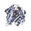
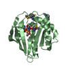
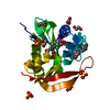
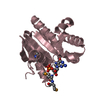
| Title | CRYSTAL STRUCTURE OF TABTOXIN RESISTANCE PROTEIN COMPLEXED WITH AN ACYL COENZYME A | ||||||
 Components Components | ACETYLTRANSFERASE | ||||||
 Keywords Keywords | TRANSFERASE / Acyl Coenzyme A complex | ||||||
| Function / homology |  Function and homology information Function and homology informationacyltransferase activity, transferring groups other than amino-acyl groups / Transferases; Acyltransferases; Transferring groups other than aminoacyl groups Similarity search - Function | ||||||
| Biological species |  Pseudomonas syringae pv. tabaci (bacteria) Pseudomonas syringae pv. tabaci (bacteria) | ||||||
| Method |  X-RAY DIFFRACTION / SYNCHROTRON / Resolution: 1.55 Å X-RAY DIFFRACTION / SYNCHROTRON / Resolution: 1.55 Å | ||||||
 Authors Authors | He, H. / Ding, Y. / Bartlam, M. / Sun, F. / Le, Y. / Qin, X. / Tang, H. / Zhang, R. / Joachimiak, A. / Liu, Y. ...He, H. / Ding, Y. / Bartlam, M. / Sun, F. / Le, Y. / Qin, X. / Tang, H. / Zhang, R. / Joachimiak, A. / Liu, Y. / Zhao, N. / Rao, Z. | ||||||
 Citation Citation | Journal: J.Mol.Biol. / Year: 2003 Title: Crystal Structure of Tabtoxin Resistance Protein Complexed with Acetyl Coenzyme A Reveals the Mechanism for beta-Lactam Acetylation Authors: He, H. / Ding, Y. / Bartlam, M. / Sun, F. / Le, Y. / Qin, X. / Tang, H. / Zhang, R. / Joachimiak, A. / Liu, Y. / Zhao, N. / Rao, Z. | ||||||
| History |
|
- Structure visualization
| Structure viewer | Molecule: MolmilJmol/JSmol |
|---|
- Downloads & links
-Download
| PDBx/mmCIF format | 1ghe.cif.gz | 82.7 KB | Display | PDBx/mmCIF format |
|---|---|---|---|---|
| PDB format | pdb1ghe.ent.gz | 62.3 KB | Display | PDB format |
| PDBx/mmJSON format | 1ghe.json.gz | Tree view | PDBx/mmJSON format | |
| Others |  Other downloads Other downloads |
-Validation report
| Arichive directory | https://data.pdbj.org/pub/pdb/validation_reports/gh/1gheftp://data.pdbj.org/pub/pdb/validation_reports/gh/1ghe | HTTPS FTP |
|---|
-Related structure data


-Links
 PDBj
PDBj

- Assembly
| Deposited unit | 
| ||||||||
|---|---|---|---|---|---|---|---|---|---|
| 1 | 
| ||||||||
| 2 | 
| ||||||||
| Unit cell |
|
-Components
| #1: Protein | Mass: 19492.443 Da / Num. of mol.: 2 Source method: isolated from a genetically manipulated source Source: (gene. exp.) Pseudomonas syringae pv. tabaci (bacteria)Species: Pseudomonas amygdali / Plasmid: PQE-30 / Production host: References: UniProt: P16966, Transferases; Acyltransferases; Transferring groups other than aminoacyl groups #2: Chemical |   Mass: 809.571 Da / Num. of mol.: 2 / Source method: obtained synthetically / Formula: C23H38N7O17P3S Mass: 809.571 Da / Num. of mol.: 2 / Source method: obtained synthetically / Formula: C23H38N7O17P3S#3: Water | ChemComp-HOH / |  Mass: 18.015 Da / Num. of mol.: 162 / Source method: isolated from a natural source / Formula: H2O Mass: 18.015 Da / Num. of mol.: 162 / Source method: isolated from a natural source / Formula: H2OHas protein modification | Y | |
|---|
-Experimental details
-Experiment
| Experiment | Method: X-RAY DIFFRACTION / Number of used crystals: 1 |
|---|
- Sample preparation
| Crystal | Density Matthews: 2.42 Å3/Da / Density % sol: 49.1 % | ||||||||||||||||||||||||||||||||||||||||||||||||||||||||||||||||||||||
|---|---|---|---|---|---|---|---|---|---|---|---|---|---|---|---|---|---|---|---|---|---|---|---|---|---|---|---|---|---|---|---|---|---|---|---|---|---|---|---|---|---|---|---|---|---|---|---|---|---|---|---|---|---|---|---|---|---|---|---|---|---|---|---|---|---|---|---|---|---|---|---|
| Crystal grow | Temperature: 291 K / Method: hanging drop/vapor diffusion / pH: 8 Details: PEG 4000, sodium acetate, Tris-HCL, pH 8.0, HANGING DROP/VAPOR DIFFUSION, temperature 291.0K | ||||||||||||||||||||||||||||||||||||||||||||||||||||||||||||||||||||||
| Crystal grow | *PLUS Temperature: 291 K / Method: vapor diffusion, hanging drop | ||||||||||||||||||||||||||||||||||||||||||||||||||||||||||||||||||||||
| Components of the solutions | *PLUS
|
-Data collection
| Diffraction | Mean temperature: 115 K |
|---|---|
| Diffraction source | Source: SYNCHROTRON / Site: APS  / Beamline: 19-ID / Wavelength: 0.9639 / Beamline: 19-ID / Wavelength: 0.9639 |
| Detector | Type: SBC-2 / Detector: CCD / Date: Sep 1, 2000 |
| Radiation | Protocol: SINGLE WAVELENGTH / Monochromatic (M) / Laue (L): M / Scattering type: x-ray |
| Radiation wavelength | Wavelength: 0.9639 Å / Relative weight: 1 |
| Reflection | Resolution: 1.4→100 Å / Num. all: 105976 / Num. obs: 93060 / % possible obs: 95.3 % / Observed criterion σ(I): -3 / Redundancy: 3 % / Biso Wilson estimate: 13.4 Å2 / Rmerge(I) obs: 0.067 / Net I/σ(I): 5.9 |
| Reflection shell | Resolution: 1.4→1.45 Å / Redundancy: 1 % / Rmerge(I) obs: 0.32 / Num. unique all: 4747 / % possible all: 65 |
| Reflection | *PLUS Highest resolution: 1.49 Å / Lowest resolution: 100 Å / Num. obs: 56060 / % possible obs: 92 % / Num. measured all: 307917 / Rmerge(I) obs: 0.071 |
| Reflection shell | *PLUS Mean I/σ(I) obs: 2.5 |
- Processing
| Software |
| |||||||||||||||||||||
|---|---|---|---|---|---|---|---|---|---|---|---|---|---|---|---|---|---|---|---|---|---|---|
| Refinement | Resolution: 1.55→30 Å / σ(F): 0 / σ(I): 0 / Stereochemistry target values: Engh & Huber / Details: CNS
| |||||||||||||||||||||
| Refinement step | Cycle: LAST / Resolution: 1.55→30 Å
| |||||||||||||||||||||
| Refine LS restraints |
| |||||||||||||||||||||
| Refinement | *PLUS Lowest resolution: 30 Å / % reflection Rfree: 10 % / Rfactor Rfree: 0.23 | |||||||||||||||||||||
| Solvent computation | *PLUS | |||||||||||||||||||||
| Displacement parameters | *PLUS | |||||||||||||||||||||
| Refine LS restraints | *PLUS
|
