 Movie
Movie Controller

[English] 日本語
 Yorodumi
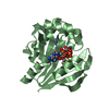
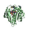
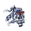
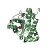
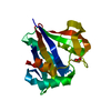
- PDB-3gxk: The crystal structure of g-type lysozyme from Atlantic cod (Gadus... -
+ Open data

- Basic information
| Entry | Database: PDB / ID: 3gxk | ||||||
|---|---|---|---|---|---|---|---|
| Title | The crystal structure of g-type lysozyme from Atlantic cod (Gadus morhua L.) in complex with NAG oligomers sheds new light on substrate binding and the catalytic mechanism. Native structure to 1.9 | ||||||
 Components Components | Goose-type lysozyme 1 | ||||||
 Keywords Keywords | HYDROLASE / Atlantic cod / fish lysozyme / active site residues / substrate binding sites / surface potential / muramidase activity / immune system | ||||||
| Function / homology |  Function and homology information Function and homology informationpeptidoglycan catabolic process / lysozyme / lysozyme activity / killing of cells of another organism / defense response to Gram-positive bacterium / extracellular region Similarity search - Function | ||||||
| Biological species |  Gadus morhua (Atlantic cod) Gadus morhua (Atlantic cod) | ||||||
| Method |  X-RAY DIFFRACTION / SYNCHROTRON / MOLECULAR REPLACEMENT / Resolution: 1.9 Å X-RAY DIFFRACTION / SYNCHROTRON / MOLECULAR REPLACEMENT / Resolution: 1.9 Å | ||||||
 Authors Authors | Helland, R. / Larsen, R.L. / Finstad, S. / Kyomuhendo, P. / Larsen, A.N. | ||||||
 Citation Citation | Journal: Cell.Mol.Life Sci. / Year: 2009 Title: Crystal structures of g-type lysozyme from Atlantic cod shed new light on substrate binding and the catalytic mechanism. Authors: Helland, R. / Larsen, R.L. / Finstad, S. / Kyomuhendo, P. / Larsen, A.N. | ||||||
| History |
|
- Structure visualization
| Structure viewer | Molecule: MolmilJmol/JSmol |
|---|
- Downloads & links
-Download
| PDBx/mmCIF format | 3gxk.cif.gz | 154 KB | Display | PDBx/mmCIF format |
|---|---|---|---|---|
| PDB format | pdb3gxk.ent.gz | 123.2 KB | Display | PDB format |
| PDBx/mmJSON format | 3gxk.json.gz | Tree view | PDBx/mmJSON format | |
| Others |  Other downloads Other downloads |
-Validation report
| Arichive directory | https://data.pdbj.org/pub/pdb/validation_reports/gx/3gxkftp://data.pdbj.org/pub/pdb/validation_reports/gx/3gxk | HTTPS FTP |
|---|
-Related structure data
| Related structure data |  3gxrC  154lS C: citing same article ( S: Starting model for refinement |
|---|---|
| Similar structure data |


-Links
 PDBj
PDBj
- Assembly
| Deposited unit | 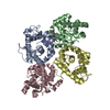
| ||||||||
|---|---|---|---|---|---|---|---|---|---|
| 1 | 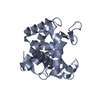
| ||||||||
| 2 | 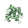
| ||||||||
| 3 | 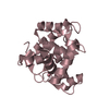
| ||||||||
| 4 | 
| ||||||||
| Unit cell |
| ||||||||
| Components on special symmetry positions |
|
-Components
| #1: Protein | Mass: 20831.008 Da / Num. of mol.: 4 / Fragment: UNP residues 31-217 Source method: isolated from a genetically manipulated source Source: (gene. exp.) Gadus morhua (Atlantic cod) / Gene: lysozyme / Plasmid: pET151/D-TOPO gLYS / Production host:  #2: Chemical |   Mass: 58.933 Da / Num. of mol.: 2 / Source method: obtained synthetically / Formula: Co Mass: 58.933 Da / Num. of mol.: 2 / Source method: obtained synthetically / Formula: Co#3: Water | ChemComp-HOH / |  Mass: 18.015 Da / Num. of mol.: 257 / Source method: isolated from a natural source / Formula: H2O Mass: 18.015 Da / Num. of mol.: 257 / Source method: isolated from a natural source / Formula: H2OSequence details | THE DISCREPANC | |
|---|
-Experimental details
-Experiment
| Experiment | Method: X-RAY DIFFRACTION / Number of used crystals: 1 |
|---|
- Sample preparation
| Crystal | Density Matthews: 1.95 Å3/Da / Density % sol: 36.91 % |
|---|---|
| Crystal grow | Temperature: 298 K / Method: vapor diffusion, hanging drop / pH: 6.5 Details: 48-53% ammonium sulphate, 5mM CoCl2, 0.1M BisTris, pH 6.5, VAPOR DIFFUSION, HANGING DROP, temperature 298K |
-Data collection
| Diffraction | Mean temperature: 100 K |
|---|---|
| Diffraction source | Source: SYNCHROTRON / Site: BESSY  / Beamline: 14.1 / Wavelength: 0.91841 Å / Beamline: 14.1 / Wavelength: 0.91841 Å |
| Detector | Type: MARMOSAIC 225 mm CCD / Detector: CCD / Date: Dec 4, 2007 |
| Radiation | Protocol: SINGLE WAVELENGTH / Monochromatic (M) / Laue (L): M / Scattering type: x-ray |
| Radiation wavelength | Wavelength: 0.91841 Å / Relative weight: 1 |
| Reflection | Resolution: 1.9→48.02 Å / Num. all: 50454 / Num. obs: 50202 / % possible obs: 99.5 % / Observed criterion σ(I): 2 / Redundancy: 4.5 % / Biso Wilson estimate: 18.99 Å2 / Rmerge(I) obs: 0.06 / Rsym value: 0.06 / Net I/σ(I): 9.4 |
| Reflection shell | Resolution: 1.9→2 Å / Redundancy: 4.3 % / Rmerge(I) obs: 0.221 / Mean I/σ(I) obs: 3.3 / Num. unique all: 7068 / Rsym value: 0.221 / % possible all: 96.9 |
- Processing
| Software |
| ||||||||||||||||||||||||||||||||||||||||||||||||||||||||||||||||||||||||||||||||||||||||||
|---|---|---|---|---|---|---|---|---|---|---|---|---|---|---|---|---|---|---|---|---|---|---|---|---|---|---|---|---|---|---|---|---|---|---|---|---|---|---|---|---|---|---|---|---|---|---|---|---|---|---|---|---|---|---|---|---|---|---|---|---|---|---|---|---|---|---|---|---|---|---|---|---|---|---|---|---|---|---|---|---|---|---|---|---|---|---|---|---|---|---|---|
| Refinement | Method to determine structure: MOLECULAR REPLACEMENT Starting model: 154L Resolution: 1.9→48.02 Å / Cor.coef. Fo:Fc: 0.939 / Cor.coef. Fo:Fc free: 0.896 / SU B: 3.827 / SU ML: 0.116 / Cross valid method: THROUGHOUT / ESU R: 0.186 / ESU R Free: 0.173 / Stereochemistry target values: MAXIMUM LIKELIHOOD / Details: HYDROGENS HAVE BEEN ADDED IN THE RIDING POSITIONS
| ||||||||||||||||||||||||||||||||||||||||||||||||||||||||||||||||||||||||||||||||||||||||||
| Solvent computation | Ion probe radii: 0.8 Å / Shrinkage radii: 0.8 Å / VDW probe radii: 1.2 Å / Solvent model: MASK | ||||||||||||||||||||||||||||||||||||||||||||||||||||||||||||||||||||||||||||||||||||||||||
| Displacement parameters | Biso mean: 18.764 Å2
| ||||||||||||||||||||||||||||||||||||||||||||||||||||||||||||||||||||||||||||||||||||||||||
| Refinement step | Cycle: LAST / Resolution: 1.9→48.02 Å
| ||||||||||||||||||||||||||||||||||||||||||||||||||||||||||||||||||||||||||||||||||||||||||
| Refine LS restraints |
| ||||||||||||||||||||||||||||||||||||||||||||||||||||||||||||||||||||||||||||||||||||||||||
| LS refinement shell | Resolution: 1.901→1.95 Å / Total num. of bins used: 20
|
