 Movie
Movie Controller

[English] 日本語
 Yorodumi
- PDB-1g1t: CRYSTAL STRUCTURE OF E-SELECTIN LECTIN/EGF DOMAINS COMPLEXED WITH SLEX -
+ Open data

- Basic information
| Entry | Database: PDB / ID: 1g1t | |||||||||
|---|---|---|---|---|---|---|---|---|---|---|
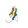
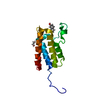
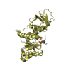
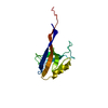
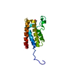
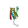
| Title | CRYSTAL STRUCTURE OF E-SELECTIN LECTIN/EGF DOMAINS COMPLEXED WITH SLEX | |||||||||
 Components Components | E-SELECTIN | |||||||||
 Keywords Keywords | IMMUNE SYSTEM / MEMBRANE PROTEIN / Lectin / EGF / Adhesion molecule / SLeX | |||||||||
| Function / homology |  Function and homology information Function and homology informationactin filament-based process / positive regulation of leukocyte tethering or rolling / sialic acid binding / leukocyte migration involved in inflammatory response / oligosaccharide binding / leukocyte tethering or rolling / positive regulation of leukocyte migration / heterophilic cell-cell adhesion / leukocyte cell-cell adhesion / cortical cytoskeleton ...actin filament-based process / positive regulation of leukocyte tethering or rolling / sialic acid binding / leukocyte migration involved in inflammatory response / oligosaccharide binding / leukocyte tethering or rolling / positive regulation of leukocyte migration / heterophilic cell-cell adhesion / leukocyte cell-cell adhesion / cortical cytoskeleton / positive regulation of receptor internalization / response to tumor necrosis factor / phospholipase binding / clathrin-coated pit / response to cytokine / response to interleukin-1 / Cell surface interactions at the vascular wall / calcium-mediated signaling / caveola / transmembrane signaling receptor activity / regulation of inflammatory response / response to lipopolysaccharide / phospholipase C-activating G protein-coupled receptor signaling pathway / membrane raft / inflammatory response / external side of plasma membrane / perinuclear region of cytoplasm / : / metal ion binding / plasma membrane Similarity search - Function | |||||||||
| Biological species |  Homo sapiens (human) Homo sapiens (human) | |||||||||
| Method |  X-RAY DIFFRACTION / MOLECULAR REPLACEMENT / Resolution: 1.5 Å X-RAY DIFFRACTION / MOLECULAR REPLACEMENT / Resolution: 1.5 Å | |||||||||
 Authors Authors | Somers, W.S. / Camphausen, R.T. | |||||||||
 Citation Citation | Journal: Cell(Cambridge,Mass.) / Year: 2000 Title: Insights into the molecular basis of leukocyte tethering and rolling revealed by structures of P- and E-selectin bound to SLe(X) and PSGL-1. Authors: Somers, W.S. / Tang, J. / Shaw, G.D. / Camphausen, R.T. | |||||||||
| History |
|
- Structure visualization
| Structure viewer | Molecule: MolmilJmol/JSmol |
|---|
- Downloads & links
-Download
| PDBx/mmCIF format | 1g1t.cif.gz | 53.1 KB | Display | PDBx/mmCIF format |
|---|---|---|---|---|
| PDB format | pdb1g1t.ent.gz | 36.1 KB | Display | PDB format |
| PDBx/mmJSON format | 1g1t.json.gz | Tree view | PDBx/mmJSON format | |
| Others |  Other downloads Other downloads |
-Validation report
| Arichive directory | https://data.pdbj.org/pub/pdb/validation_reports/g1/1g1tftp://data.pdbj.org/pub/pdb/validation_reports/g1/1g1t | HTTPS FTP |
|---|
-Related structure data


-Links
 PDBj
PDBj


- Assembly
| Deposited unit | 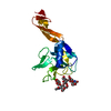
| ||||||||||
|---|---|---|---|---|---|---|---|---|---|---|---|
| 1 |
| ||||||||||
| Unit cell |
|
-Components
| #1: Protein | Mass: 18103.234 Da / Num. of mol.: 1 / Fragment: LECTIN/EGF DOMAINS Source method: isolated from a genetically manipulated source Source: (gene. exp.) Homo sapiens (human) / Cell (production host): ovary [CHO] cells / Production host:   Cricetulus griseus (Chinese hamster) / References: UniProt: P16581 Cricetulus griseus (Chinese hamster) / References: UniProt: P16581 |
|---|---|
| #2: Polysaccharide | N-acetyl-alpha-neuraminic acid-(2-3)-beta-D-galactopyranose-(1-4)-[alpha-L-fucopyranose-(1-3)] ...N-acetyl-alpha-neuraminic acid-(2-3)-beta-D-galactopyranose-(1-4)-[alpha-L-fucopyranose-(1-3)]methyl 2-acetamido-2-deoxy-beta-D-glucopyranoside Source method: isolated from a genetically manipulated source |
| #3: Chemical | ChemComp-CA /   Mass: 40.078 Da / Num. of mol.: 1 / Source method: obtained synthetically / Formula: Ca Mass: 40.078 Da / Num. of mol.: 1 / Source method: obtained synthetically / Formula: Ca |
| #4: Water | ChemComp-HOH /  Mass: 18.015 Da / Num. of mol.: 186 / Source method: isolated from a natural source / Formula: H2O Mass: 18.015 Da / Num. of mol.: 186 / Source method: isolated from a natural source / Formula: H2O |
| Has protein modification | Y |
-Experimental details
-Experiment
| Experiment | Method: X-RAY DIFFRACTION / Number of used crystals: 1 |
|---|
- Sample preparation
| Crystal | Density Matthews: 2.68 Å3/Da / Density % sol: 54.02 % | ||||||||||||||||||||||||||||||||||||
|---|---|---|---|---|---|---|---|---|---|---|---|---|---|---|---|---|---|---|---|---|---|---|---|---|---|---|---|---|---|---|---|---|---|---|---|---|---|
| Crystal grow | Temperature: 291 K / Method: vapor diffusion, hanging drop / pH: 7.5 Details: HEPES, Tris-HCl, CaCl2, PEG 4000, pH 7.5, VAPOR DIFFUSION, HANGING DROP at 291K | ||||||||||||||||||||||||||||||||||||
| Crystal grow | *PLUS Temperature: 18 ℃ / Method: vapor diffusion | ||||||||||||||||||||||||||||||||||||
| Components of the solutions | *PLUS
|
-Data collection
| Diffraction | Mean temperature: 100 K |
|---|---|
| Diffraction source | Source: ROTATING ANODE / Type: RIGAKU RU200 / Wavelength: 1.5418 |
| Detector | Type: RIGAKU RAXIS IV / Detector: IMAGE PLATE / Date: Jan 1, 1997 / Details: Mirrors |
| Radiation | Monochromator: Mirrors / Protocol: SINGLE WAVELENGTH / Monochromatic (M) / Laue (L): M / Scattering type: x-ray |
| Radiation wavelength | Wavelength: 1.5418 Å / Relative weight: 1 |
| Reflection | Resolution: 1.5→15 Å / Num. all: 29493 / Num. obs: 29493 / % possible obs: 92.4 % / Observed criterion σ(I): 3 / Redundancy: 8.8 % / Biso Wilson estimate: 20 Å2 / Rmerge(I) obs: 0.041 / Net I/σ(I): 47.6 |
| Reflection shell | Resolution: 1.5→1.55 Å / Redundancy: 8.8 % / Rmerge(I) obs: 0.22 / % possible all: 62 |
| Reflection | *PLUS Lowest resolution: 15 Å / Num. measured all: 260283 |
| Reflection shell | *PLUS % possible obs: 62 % / Rmerge(I) obs: 0.22 / Mean I/σ(I) obs: 4.4 |
- Processing
| Software |
| |||||||||||||||||||||||||
|---|---|---|---|---|---|---|---|---|---|---|---|---|---|---|---|---|---|---|---|---|---|---|---|---|---|---|
| Refinement | Method to determine structure: MOLECULAR REPLACEMENT / Resolution: 1.5→15 Å / Cross valid method: THROUGHOUT / σ(F): 0 / σ(I): 0 / Stereochemistry target values: Engh & Huber
| |||||||||||||||||||||||||
| Refinement step | Cycle: LAST / Resolution: 1.5→15 Å
| |||||||||||||||||||||||||
| Refine LS restraints |
| |||||||||||||||||||||||||
| Software | *PLUS Name: CNS / Classification: refinement | |||||||||||||||||||||||||
| Refinement | *PLUS Lowest resolution: 15 Å / σ(F): 0 / % reflection Rfree: 5 % | |||||||||||||||||||||||||
| Solvent computation | *PLUS | |||||||||||||||||||||||||
| Displacement parameters | *PLUS |
