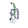
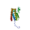
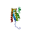
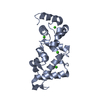
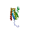
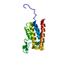
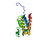
: / ameboidal-type cell migration / Amplification of signal from unattached kinetochores via a MAD2 inhibitory signal / COPI-independent Golgi-to-ER retrograde traffic / Resolution of Sister Chromatid Cohesion / Mitotic Prometaphase / EML4 and NUDC in mitotic spindle formation / RHO GTPases Activate Formins / platelet-activating factor acetyltransferase activity / Loss of Nlp from mitotic centrosomes ...: / ameboidal-type cell migration / Amplification of signal from unattached kinetochores via a MAD2 inhibitory signal / COPI-independent Golgi-to-ER retrograde traffic / Resolution of Sister Chromatid Cohesion / Mitotic Prometaphase / EML4 and NUDC in mitotic spindle formation / RHO GTPases Activate Formins / platelet-activating factor acetyltransferase activity / Loss of Nlp from mitotic centrosomes / Recruitment of mitotic centrosome proteins and complexes / Loss of proteins required for interphase microtubule organization from the centrosome / Recruitment of NuMA to mitotic centrosomes / Anchoring of the basal body to the plasma membrane / AURKA Activation by TPX2 / maintenance of centrosome location / Regulation of PLK1 Activity at G2/M Transition / Separation of Sister Chromatids / microtubule cytoskeleton organization involved in establishment of planar polarity / establishment of planar polarity of embryonic epithelium / 1-alkyl-2-acetylglycerophosphocholine esterase complex / COPI-independent Golgi-to-ER retrograde traffic / corpus callosum morphogenesis / cerebral cortex neuron differentiation / positive regulation of cytokine-mediated signaling pathway / Amplification of signal from unattached kinetochores via a MAD2 inhibitory signal / establishment of centrosome localization / acrosome assembly / central region of growth cone / nuclear membrane disassembly / auditory receptor cell development / microtubule sliding / positive regulation of cellular component organization / Mitotic Prometaphase / EML4 and NUDC in mitotic spindle formation / Resolution of Sister Chromatid Cohesion / positive regulation of embryonic development / microtubule organizing center organization / layer formation in cerebral cortex / osteoclast development / RHO GTPases Activate Formins / Loss of Nlp from mitotic centrosomes / Recruitment of mitotic centrosome proteins and complexes / Loss of proteins required for interphase microtubule organization from the centrosome / Anchoring of the basal body to the plasma membrane / Separation of Sister Chromatids / Recruitment of NuMA to mitotic centrosomes / AURKA Activation by TPX2 / astral microtubule / cortical microtubule organization / Regulation of PLK1 Activity at G2/M Transition / reelin-mediated signaling pathway / positive regulation of dendritic spine morphogenesis / brain morphogenesis / stereocilium / microtubule plus-end binding / regulation of GTPase activity / motile cilium / stem cell division / vesicle transport along microtubule / retrograde axonal transport / negative regulation of JNK cascade / neuromuscular process controlling balance / regulation of postsynapse organization / microtubule associated complex / kinesin complex / nuclear migration / establishment of mitotic spindle orientation / dynein intermediate chain binding / neuroblast proliferation / germ cell development / cell leading edge / transmission of nerve impulse / dynein complex binding / protein secretion / cochlea development / lipid catabolic process / positive regulation of axon extension / microtubule-based process / cytoplasmic microtubule / positive regulation of mitotic cell cycle / adult locomotory behavior / axon cytoplasm / hippocampus development / regulation of microtubule cytoskeleton organization / phosphoprotein binding / brain development / cerebral cortex development / microtubule cytoskeleton organization / modulation of chemical synaptic transmission / neuron migration / kinetochore / Schaffer collateral - CA1 synapse / nuclear envelope / negative regulation of neuron projection development / cell migration / sperm midpiece / microtubule cytoskeleton / growth cone / actin cytoskeleton organization Similarity search - Function
Mass: 18.015 Da / Num. of mol.: 181 / Source method: isolated from a natural source / Formula: H2O
Has protein modification
Y
Sequence details
INITIAL 2 RESIDUES ARE CLONING ARTIFACTS.
-
Experimental details
-
Experiment
Experiment
Method: X-RAY DIFFRACTION / Number of used crystals: 1
-
Sample preparation
Crystal
Density Matthews: 2.1 Å3/Da / Density % sol: 49.6 %
Crystal grow
Method: microbatch / pH: 4.5 Details: CRYSTALS WERE GROWN USING SITTING-DROP VAPOUR-DIFFUSION UNDER MINERAL OIL USING A 1:1 MIXTURE OF PROTEIN AND 1.7 M (NH4)2SO4 AND 0.1 M NA3-CITRATE, PH 4.5
 Movie
Movie Controller


 Open data
 Basic information
 Components
 Keywords
 Function and homology information
 X-RAY DIFFRACTION /
  Authors
 Citation
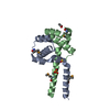
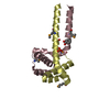
 Structure visualization
 Downloads & links
 Other downloads
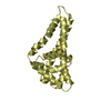

 PDBj
PDBj


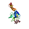
 Assembly
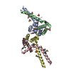


 Mass: 96.063 Da / Num. of mol.: 4 / Source method: obtained synthetically / Formula: SO4
 Mass: 59.044 Da / Num. of mol.: 1 / Source method: obtained synthetically / Formula: C2H3O2
 Mass: 122.121 Da / Num. of mol.: 1 / Source method: obtained synthetically / Formula: C7H6O2
 Mass: 18.015 Da / Num. of mol.: 181 / Source method: isolated from a natural source / Formula: H2O
 Sample preparation
 / Beamline: 22-ID / Wavelength: 0.979392,0.979528, 0.964216
 Processing